Explore
 Validate
 Learn
Learn Western blot
Antibody data
- Antibody Data
- Antigen structure
- References [3]
- Comments [0]
- Validations
- Western blot [2]
- Immunohistochemistry [1]
Submit
Validation data
Reference
Comment
Report error
- Product number
- AF3156 - Provider product page

- Provider
- R&D Systems
- Product name
- Human Serpin A8/Angiotensinogen Antibody
- Antibody type
- Polyclonal
- Description
- Immunogen affinity purified. Detects human Serpin A8/Angiotensinogen in direct ELISAs and Western blots. In direct ELISAs, approximately 20% cross-reactivity with recombinant mouse Serpin A8/Angiotensinogen is observed, and less than 5% cross-reactivity with recombinant human Serpin A1, A4, A5, F2, G1, I1, and I2 is observed.
- Reactivity
- Human
- Host
- Goat
- Conjugate
- Unconjugated
- Antigen sequence
ABM85122- Isotype
- IgG
- Vial size
- 100 ug
- Concentration
- LYOPH
- Storage
- Use a manual defrost freezer and avoid repeated freeze-thaw cycles. 12 months from date of receipt, -20 to -70 °C as supplied. 1 month, 2 to 8 °C under sterile conditions after reconstitution. 6 months, -20 to -70 °C under sterile conditions after reconstitution.
Submitted references Expression of renin-angiotensin system (RAS) components in endometrial cancer.
Hepatocarcinogenesis driven by GSNOR deficiency is prevented by iNOS inhibition.
The inflammatory response in the MPTP model of Parkinson's disease is mediated by brain angiotensin: relevance to progression of the disease.
Delforce SJ, Lumbers ER, Corbisier de Meaultsart C, Wang Y, Proietto A, Otton G, Scurry J, Verrills NM, Scott RJ, Pringle KG
Endocrine connections 2017 Jan;6(1):9-19
Endocrine connections 2017 Jan;6(1):9-19
Hepatocarcinogenesis driven by GSNOR deficiency is prevented by iNOS inhibition.
Tang CH, Wei W, Hanes MA, Liu L
Cancer research 2013 May 1;73(9):2897-904
Cancer research 2013 May 1;73(9):2897-904
The inflammatory response in the MPTP model of Parkinson's disease is mediated by brain angiotensin: relevance to progression of the disease.
Joglar B, Rodriguez-Pallares J, Rodriguez-Perez AI, Rey P, Guerra MJ, Labandeira-Garcia JL
Journal of neurochemistry 2009 Apr;109(2):656-69
Journal of neurochemistry 2009 Apr;109(2):656-69
No comments: Submit comment
Supportive validation
- Submitted by
- R&D Systems (provider)
- Main image

- Experimental details
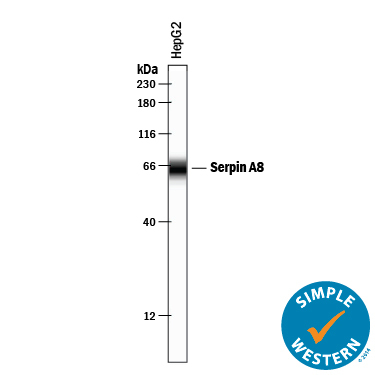
- Detection of Human Serpin A8/Angiotensinogen by Simple WesternTM. Simple Western lane view shows lysates of HepG2 human hepatocellular carcinoma cell line, loaded at 0.2 mg/mL. A specific band was detected for Serpin A8/Angiotensinogen at approximately 64 kDa (as indicated) using 10 µg/mL of Goat Anti-Human Serpin A8/Angiotensinogen Antigen Affinity-purified Polyclonal Antibody (Catalog # AF3156) followed by 1:50 dilution of HRP-conjugated Anti-Goat IgG Secondary Antibody (Catalog # HAF109). This experiment was conducted under reducing conditions and using the 12-230 kDa separation system.
- Submitted by
- R&D Systems (provider)
- Main image
- Experimental details
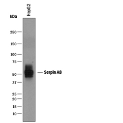
- Detection of Human Serpin A8/Angiotensinogen by Western Blot. Western blot shows lysates of HepG2 human hepatocellular carcinoma cell line. PVDF membrane was probed with 1 µg/mL of Goat Anti-Human Serpin A8/Angiotensinogen Antigen Affinity-purified Polyclonal Antibody (Catalog # AF3156) followed by HRP-conjugated Anti-Goat IgG Secondary Antibody (Catalog # HAF019). A specific band was detected for Serpin A8/Angiotensinogen at approximately 50-55 kDa (as indicated). This experiment was conducted under reducing conditions and using Immunoblot Buffer Group 1.

Supportive validation
- Submitted by
- R&D Systems (provider)
- Main image

- Experimental details
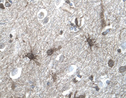
- Serpin A8/Angiotensinogen in Human Brain. Serpin A8/Angiotensinogen was detected in immersion fixed paraffin-embedded sections of human brain (cortex) using 1.7 µg/mL Goat Anti-Human Serpin A8/Angiotensinogen Antigen Affinity-purified Polyclonal Antibody (Catalog # AF3156) overnight at 4 °C. Tissue was stained with the Anti-Goat HRP-DAB Cell & Tissue Staining Kit (brown; Catalog # CTS008) and counterstained with hematoxylin (blue). View our protocol for Chromogenic IHC Staining of Paraffin-embedded Tissue Sections.