Explore
 Validate
 Learn
Learn Western blot
 ELISA
ELISAAntibody data
- Antibody Data
- Antigen structure
- References [0]
- Comments [0]
- Validations
- Western blot [2]
- Immunohistochemistry [10]
Submit
Validation data
Reference
Comment
Report error
- Product number
- LS-C782225 - Provider product page

- Provider
- LSBio
- Product name
- Lumican Antibody LS-C782225
- Antibody type
- Polyclonal
- Description
- Antigen Affinity purification
- Reactivity
- Human, Mouse, Rat
- Host
- Rabbit
- Isotype
- IgG
- Storage
- After reconstitution, store at 4°C for up to 1 month. Long-term: aliquot and store at -20°C. Avoid freeze-thaws cycles.
No comments: Submit comment
Enhanced validation
- Submitted by
- LSBio (provider)
- Enhanced method
- Genetic validation
- Main image
- Experimental details
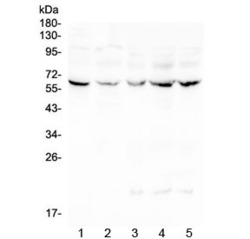
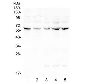
- Western blot testing of human 1) placenta, 2) Caco-2, 3) CCRF-CEM, 4) HeLa and 5) Jurkat lysate with LUM antibody at 0.5ug/ml. Expected moleculer weight: ~40 kDa (unmodified), ~60 kDa (glycosylated).
- Submitted by
- LSBio (provider)
- Enhanced method
- Genetic validation
- Main image
- Experimental details
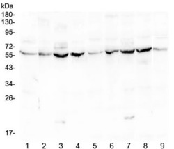
- Western blot testing of mouse 1) liver, 2) ovary, 3) testis, 4) lung and rat 5) liver, 6) ovary, 7) testis, 8) lung and 9) heart lysate with LUM antibody at 0.5ug/ml. Expected moleculer weight: ~40 kDa (unmodified), ~60 kDa (glycosylated).

Enhanced validation
- Submitted by
- LSBio (provider)
- Enhanced method
- Genetic validation
- Main image

- Experimental details
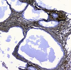
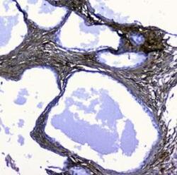
- IHC staining of FFPE human placenta with LUM antibody at 1ug/ml. HIER: boil tissue sections in pH6, 10mM citrate buffer, for 10-20 min followed by cooling at RT for 20 min.

- Submitted by
- LSBio (provider)
- Enhanced method
- Genetic validation
- Main image

- Experimental details
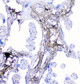
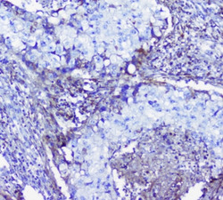
- IHC staining of FFPE human ovarian cancer with LUM antibody at 1ug/ml. HIER: boil tissue sections in pH6, 10mM citrate buffer, for 10-20 min followed by cooling at RT for 20 min.

- Submitted by
- LSBio (provider)
- Enhanced method
- Genetic validation
- Main image
- Experimental details
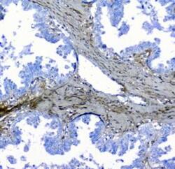
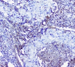
- IHC staining of FFPE human pancreatic cancer with LUM antibody at 1ug/ml. HIER: boil tissue sections in pH6, 10mM citrate buffer, for 10-20 min followed by cooling at RT for 20 min.

- Submitted by
- LSBio (provider)
- Enhanced method
- Genetic validation
- Main image

- Experimental details
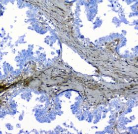
- IHC staining of FFPE human placenta with LUM antibody at 1ug/ml. HIER: boil tissue sections in pH6, 10mM citrate buffer, for 10-20 min followed by cooling at RT for 20 min.
- Submitted by
- LSBio (provider)
- Enhanced method
- Genetic validation
- Main image
- Experimental details
- IHC staining of FFPE human ovarian cancer with LUM antibody at 1ug/ml. HIER: boil tissue sections in pH6, 10mM citrate buffer, for 10-20 min followed by cooling at RT for 20 min.
- Submitted by
- LSBio (provider)
- Enhanced method
- Genetic validation
- Main image
- Experimental details
- IHC staining of FFPE human lung cancer with LUM antibody at 1ug/ml. HIER: boil tissue sections in pH6, 10mM citrate buffer, for 10-20 min followed by cooling at RT for 20 min.

- Submitted by
- LSBio (provider)
- Main image
- Experimental details
- IHC staining of FFPE human lung cancer with LUM antibody at 1ug/ml. HIER: boil tissue sections in pH6, 10mM citrate buffer, for 10-20 min followed by cooling at RT for 20 min.

- Submitted by
- LSBio (provider)
- Main image

- Experimental details
- IHC staining of FFPE human placenta with LUM antibody at 1ug/ml. HIER: boil tissue sections in pH6, 10mM citrate buffer, for 10-20 min followed by cooling at RT for 20 min.

- Submitted by
- LSBio (provider)
- Main image

- Experimental details
- IHC staining of FFPE human ovarian cancer with LUM antibody at 1ug/ml. HIER: boil tissue sections in pH6, 10mM citrate buffer, for 10-20 min followed by cooling at RT for 20 min.

- Submitted by
- LSBio (provider)
- Main image

- Experimental details
- IHC staining of FFPE human pancreatic cancer with LUM antibody at 1ug/ml. HIER: boil tissue sections in pH6, 10mM citrate buffer, for 10-20 min followed by cooling at RT for 20 min.