Explore
 Validate
 Learn
LearnGTX25478
antibody from GeneTex
Targeting: HSPD1
GroEL, HSP60, SPG13
 Western blot
 ELISA Immunocytochemistry Immunoprecipitation Immunohistochemistry Flow cytometry Blocking/Neutralizing
Antibody data
- Antibody Data
- Antigen structure
- References [0]
- Comments [0]
- Validations
- Western blot [1]
- Immunocytochemistry [1]
- Immunoprecipitation [1]
- Immunohistochemistry [1]
Submit
Validation data
Reference
Comment
Report error
- Product number
- GTX25478 - Provider product page

- Provider
- GeneTex
- Proper citation
- GeneTex Cat#GTX25478, RRID:AB_373944
- Product name
- HSP60 antibody [4B9/89]
- Antibody type
- Monoclonal
- Reactivity
- Human, Mouse, Rat, Bacteria, Simian
- Host
- Mouse
No comments: Submit comment
Supportive validation
- Submitted by
- GeneTex (provider)
- Main image
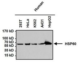
- Experimental details
- Western blot analysis of HSP60 in 50£gg of various cell lysates. Proteins were transferred to a PVDF membrane and blocked with 5% BSA/TBST for at least 1 hour. The membrane was probed with HSP60 antibody [4B9/89] at a dilution of 1:1000 overnight at 4¢XC on a rocking platform, washed in TBS-0.1%Tween 20, and probed with a proper secondary antibody. Chemiluminescent detection was performed.
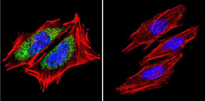
Supportive validation
- Submitted by
- GeneTex (provider)
- Main image

- Experimental details
- Immunofluorescent analysis of HSP60 in A2058 cells. HSP60 staining (green), F-Actin staining with Phalloidin (red) and nuclei with DAPI (blue) is shown. Cells were grown on slides and fixed with formaldehyde prior to staining. Cells were probed without (control) or with HSP60 antibody [4B9/89] at a dilution of 1:200 over night at 4?C, washed with PBS and incubated with a proper secondary antibody. Images were taken at 60X magnification.
Supportive validation
- Submitted by
- GeneTex (provider)
- Main image
- Experimental details
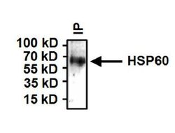
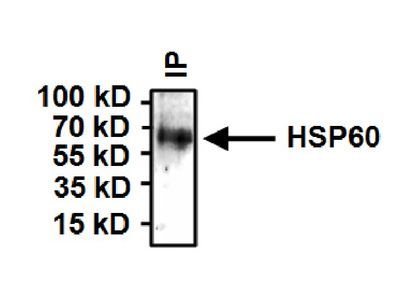
- Immunoprecipitation of HSP60 from HeLa cells. Antigen-antibody complexes were formed by incubating 500£gg whole cell lysate with 2£gg of HSP60 antibody [4B9/89] overnight on a rocking platform at 4¢XC. The immune complexes were captured on 50£gl Protein A/G Agarose , washed extensively, and eluted. Samples were then resolved on a 4-20% Tris-HCl polyacrylamide gel, transferred to a PVDF membrane, and blocked with 5% BSA/TBST for at least 1 hour. The membrane was probed with HSP60 antibody [4B9/89] at a dilution of 1:1000 overnight rotating at 4¢XC, washed in TBST, and probed with a proper secondary antibody. Chemiluminescent detection was performed.
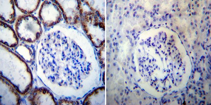
Supportive validation
- Submitted by
- GeneTex (provider)
- Main image
- Experimental details
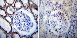
- Immunohistochemistry was performed on normal deparaffinized human Kidney tissue. To expose target proteins, heat induced antigen retrieval was performed using 10mM sodium citrate (pH6.0) buffer, microwaved for 8-15 minutes. Following antigen retrieval tissues were blocked in 3% BSA-PBS for 30 minutes at room temperature. Tissues were then probed at a dilution of 1:100 with or without HSP60 antibody [4B9/89] overnight at 4¢XC in a humidified chamber. Tissues were washed extensively with PBST and endogenous peroxidase activity was quenched with a peroxidase suppressor. Detection was performed using a biotin-conjugated secondary antibody and SA-HRP, followed by colorimetric detection using DAB. Tissues were counterstained with hematoxylin and prepped for mounting.