Explore
 Validate
 Learn
Learn Western blot
Antibody data
- Antibody Data
- Antigen structure
- References [1]
- Comments [0]
- Validations
- Western blot [6]
- Immunocytochemistry [2]
- Immunohistochemistry [6]
Submit
Validation data
Reference
Comment
Report error
- Product number
- PA5-34760 - Provider product page

- Provider
- Invitrogen Antibodies
- Product name
- HSP60 Polyclonal Antibody
- Antibody type
- Polyclonal
- Antigen
- Recombinant protein fragment
- Description
- Recommended positive controls: 293T, A431, HeLa, HepG2, mouse brain, rat brain. Predicted reactivity: Mouse (97%), Rat (97%), Zebrafish (85%), Xenopus laevis (91%), Pig (99%), Chicken (94%), Rhesus Monkey (97%), Chimpanzee (99%), Bovine (98%). Store product as a concentrated solution. Centrifuge briefly prior to opening the vial.
- Reactivity
- Human, Mouse, Rat, Hamster
- Host
- Rabbit
- Isotype
- IgG
- Vial size
- 100 µL
- Concentration
- 0.3 mg/mL
- Storage
- Store at 4°C short term. For long term storage, store at -20°C, avoiding freeze/thaw cycles.
Submitted references Heat shock protein 60 is a disease-associated sialoglycoprotein in human non-small cell lung cancer.
Singh P, Kumari M, Bal A, Srinivasan R, Ghosh S
Biological chemistry 2020 Jul 28;401(8):969-983
Biological chemistry 2020 Jul 28;401(8):969-983
No comments: Submit comment
Supportive validation
- Submitted by
- Invitrogen Antibodies (provider)
- Main image
- Experimental details
- Western blot analysis of HSP60/Heat Shock Protein 60 using 30µg of A) H1299 (B) HeLa S3 and C) MOLT4 lysate. Samples were loaded onto a 7.5% SDS-PAGE gel and probed with a HSP60/Heat Shock Protein 60 polyclonal antibody (Product # PA5-34760) at a dilution of 1:10,000.

- Submitted by
- Invitrogen Antibodies (provider)
- Main image

- Experimental details
- Western Blot analysis of HSP60 was performed by separating 30 µg of various whole cell extracts by 7.5% SDS-PAGE. Proteins were transferred to a membrane and probed with a HSP60 Polyclonal Antibody (Product # PA5-34760) at a dilution of 1:10000 and a HRP-conjugated anti-rabbit IgG secondary antibody.

- Submitted by
- Invitrogen Antibodies (provider)
- Main image

- Experimental details
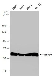
- Western blot analysis was performed on whole cell extracts (30 µg lysate) of HeLa (Lane 1), THP-1 (Lane 2), HepG2 (Lane 3), SW480 (Lane 4), COS-7 (Lane 5), NIH3T3 (Lane 6), HL-60 (Lane 7), tissue extracts of Mouse Brain (Lane 8) and Rat Brain (Lane 9). The blot was probed with Anti-HSP60 Polyclonal Antibody (Product # PA5-34760, 1:10000 dilution) and detected by chemiluminescence using Goat anti-Rabbit IgG (H+L) Superclonal™ Secondary Antibody, HRP conjugate (Product # A27036, 0.25 µg/ml, 1:4000 dilution). A 60kDa band corresponding to HSP60 was observed across all cell lines and tissues tested except HL-60.

- Submitted by
- Invitrogen Antibodies (provider)
- Main image
- Experimental details
- Western Blot analysis of HSP60 was performed by separating 30 µg of various whole cell extracts by 7.5% SDS-PAGE. Proteins were transferred to a membrane and probed with a HSP60 Polyclonal Antibody (Product # PA5-34760) at a dilution of 1:10000 and a HRP-conjugated anti-rabbit IgG secondary antibody.

- Submitted by
- Invitrogen Antibodies (provider)
- Main image

- Experimental details
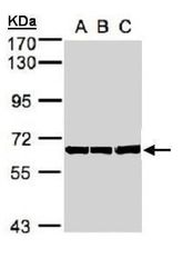
- Western Blot using HSP60 Polyclonal Antibody (Product # PA5-34760). Sample (20 µg of whole cell lysate). Lane A: mouse brain. 7.5% SDS PAGE. HSP60 Polyclonal Antibody (Product # PA5-34760) diluted at 1:20,000. The HRP-conjugated anti-rabbit IgG antibody was used to detect the primary antibody.

- Submitted by
- Invitrogen Antibodies (provider)
- Main image

- Experimental details
- HSP60 Polyclonal Antibody detects HSPD1 protein by western blot analysis. A. 50 µg rat brain lysate/extract.7.5% SDS-PAGE. HSP60 Polyclonal Antibody (Product # PA5-34760) dilution: 1:10,000. The HRP-conjugated anti-rabbit IgG antibody was used to detect the primary antibody.

Supportive validation
- Submitted by
- Invitrogen Antibodies (provider)
- Main image
- Experimental details
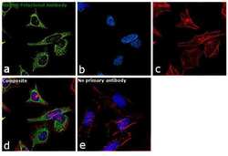
- Immunofluorescence analysis of HSP60 was performed using 70% confluent log phase HeLa cells. The cells were fixed with 4% paraformaldehyde for 10 minutes, permeabilized with 0.1% Triton™ X-100 for 15 minutes, and blocked with 1% BSA for 1 hour at room temperature. The cells were labeled with HSP60 Polyclonal Antibody (Product # PA5-34760) at 1:200 dilution in 0.1% BSA, incubated at 4 degree Celsius overnight and then labeled with Goat anti-Rabbit IgG (H+L) Superclonal™ Secondary Antibody, Alexa Fluor® 488 conjugate (Product # A27034) at a dilution of 1:2000 for 45 minutes at room temperature (Panel a: green). Nuclei (Panel b: blue) were stained with ProLong™ Diamond Antifade Mountant with DAPI (Product # P36962). F-actin (Panel c: red) was stained with Rhodamine Phalloidin (Product # R415). Panel d represents the merged image showing mitochondrial localization. Panel e represents control cells with no primary antibody to assess background. The images were captured at 60X magnification.

- Submitted by
- Invitrogen Antibodies (provider)
- Main image

- Experimental details
- Immunocytochemistry-Immunofluorescence analysis of HSP60 was performed in HeLa cells fixed in ice-cold MeOH for 5 min. Green: HSP60 Polyclonal Antibody (Product # PA5-34760) diluted at 1:500. Blue: Hoechst 33342 staining. Scale bar = 10 µm.

Supportive validation
- Submitted by
- Invitrogen Antibodies (provider)
- Main image

- Experimental details
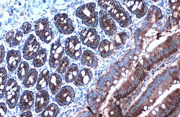
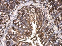
- HSP60 Polyclonal Antibody detects HSP60 protein at mitochondria by immunohistochemical analysis. Sample: Paraffin-embedded mouse intestine. HSP60 stained by HSP60 Polyclonal Antibody (Product # PA5-34760) diluted at 1:1,000. Antigen Retrieval: Citrate buffer, pH 6.0, 15 min.

- Submitted by
- Invitrogen Antibodies (provider)
- Main image

- Experimental details
- HSP60 Polyclonal Antibody detects HSP60 protein at mitochondria by immunohistochemical analysis. Sample: Paraffin-embedded rat duodenum. HSP60 stained by HSP60 Polyclonal Antibody (Product # PA5-34760) diluted at 1:500. Antigen Retrieval: Citrate buffer, pH 6.0, 15 min.
- Submitted by
- Invitrogen Antibodies (provider)
- Main image

- Experimental details
- HSP60 Polyclonal Antibody detects HSP60 protein at mitochondria by immunohistochemical analysis. Sample: Paraffin-embedded rat kidney. HSP60 stained by HSP60 Polyclonal Antibody (Product # PA5-34760) diluted at 1:1,000. Antigen Retrieval: Citrate buffer, pH 6.0, 15 min.

- Submitted by
- Invitrogen Antibodies (provider)
- Main image
- Experimental details
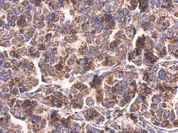
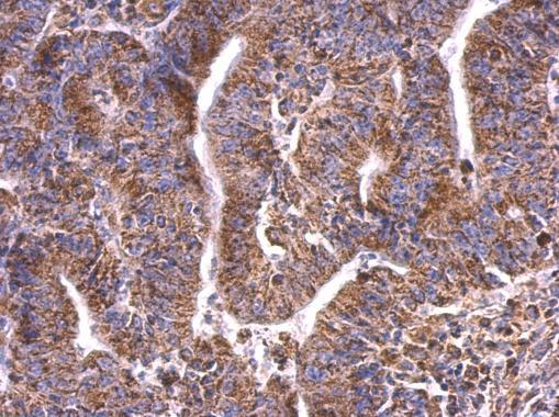
- HSP60 Polyclonal Antibody detects HSP60 protein at mitochondria on human breast carcinoma by immunohistochemical analysis. Sample: Paraffin-embedded human breast carcinoma. HSP60 Polyclonal Antibody (Product # PA5-34760) dilution: 1:500. Antigen Retrieval: EDTA based buffer, pH 8.0, 15 min.
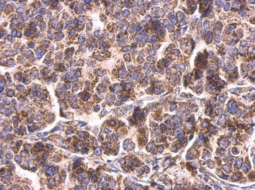
- Submitted by
- Invitrogen Antibodies (provider)
- Main image
- Experimental details
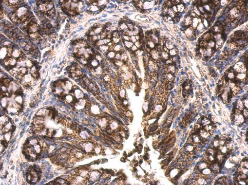
- HSP60 Polyclonal Antibody detects HSP60 protein at mitochondria on human colon carcinoma by immunohistochemical analysis. Sample: Paraffin-embedded human colon carcinoma. HSP60 Polyclonal Antibody (Product # PA5-34760) dilution: 1:500. Antigen Retrieval: EDTA based buffer, pH 8.0, 15 min.
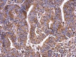
- Submitted by
- Invitrogen Antibodies (provider)
- Main image
- Experimental details
- HSP60 Polyclonal Antibody detects HSP60 protein at mitochondrion on mouse colon by immunohistochemical analysis. Sample: Paraffin-embedded mouse colon. HSP60 Polyclonal Antibody (Product # PA5-34760) dilution: 1:500. Antigen Retrieval: EDTA based buffer, pH 8.0, 15 min.