 Explore
 Validate
 Learn
Learn Western blot
 Immunocytochemistry
Antibody data
- Antibody Data
- Antigen structure
- References [0]
- Comments [0]
- Validations
- Western blot [3]
- Immunohistochemistry [2]
- Flow cytometry [2]
Submit
Validation data
Reference
Comment
Report error
- Product number
- NBP2-32973-0.1mg - Provider product page

- Provider
- Novus Biologicals
- Product name
- Mouse Monoclonal HSP60 Antibody
- Antibody type
- Monoclonal
- Description
- Protein A purified. Recognizes a 60kDa protein, identified as the heat shock protein 60 (hsp60). Its epitope is localized between aa 383-447 of human hsp60. A wide variety of environmental and pathophysiological stressful conditions trigger the synthesis of a family of proteins known as heat shock proteins (hsps), more appropriately called as stress response proteins (srps). hsp60 is a potential antigen in a number of autoimmune diseases. In human arthritis and in experimentally induced arthritis in animals, disease development coincides with the development of immune reactivity directed against not only bacterial hsp60, but also against its mammalian homolog. Clone LK1, unlike LK2, recognizes only the mammalian (not bacterial) hsp60 and is useful in distinguishing hsp60 from mammals and bacteria.
- Reactivity
- Human, Mouse, Rat, Bovine, Canine, Chicken/Avian, Drosophila, Hamster, Porcine, Rabbit, Sheep, Simian, Xenopus
- Host
- Mouse
- Isotype
- IgG
- Vial size
- 0.1 mg
- Concentration
- 0.2 mg/ml
- Storage
- Store at 4C.
No comments: Submit comment
Supportive validation
- Submitted by
- Novus Biologicals (provider)
- Main image

- Experimental details
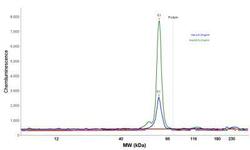
- Simple Western: HSP60 Antibody (LK1) [NBP2-32973] - Simple Western lane view shows a specific band for HSP60 in 0.2 mg/ml of HeLa (left) and HepG2 (right) lysate(s). This experiment was performed under reducing conditions using the 12-230 kDa separation system.

- Submitted by
- Novus Biologicals (provider)
- Main image
- Experimental details
- Simple Western: HSP60 Antibody (LK1) [NBP2-32973] - Electropherogram image of the corresponding Simple Western lane. HSP60 antibody was used at 10 ug/ml dilution of HeLa and HepG2 lysates(s) respectively.

- Submitted by
- Novus Biologicals (provider)
- Main image

- Experimental details
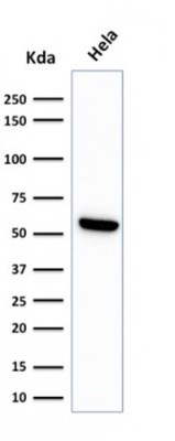
- Western Blot: HSP60 Antibody (LK1) [NBP2-32973] - Western Blot Analysis of HeLa cell lysate using HSP60 Antibody (LK1)
Supportive validation
- Submitted by
- Novus Biologicals (provider)
- Main image
- Experimental details
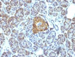
- Immunohistochemistry-Paraffin: HSP60 Antibody (LK1) [NBP2-32973] - Formalin-fixed paraffin-embedded human pancreas stained with HSP60 Monoclonal Antibody (LK1).

- Submitted by
- Novus Biologicals (provider)
- Main image

- Experimental details
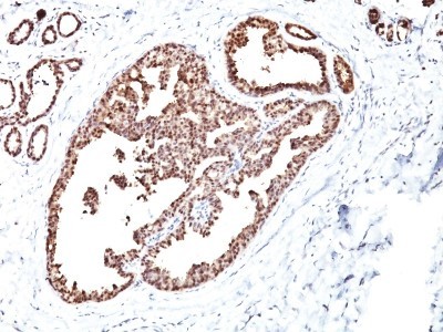
- Immunohistochemistry-Paraffin: HSP60 Antibody (LK1) [NBP2-32973] - Formalin-fixed, paraffin-embedded human breast carcinoma stained with HSP60 MAb (LK1)
Supportive validation
- Submitted by
- Novus Biologicals (provider)
- Main image

- Experimental details
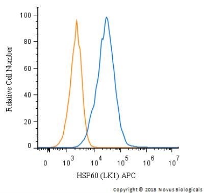
- Flow Cytometry: HSP60 Antibody (LK1) [NBP2-32973] - An intracellular stain was performed on HeLa cells with HSP60 Antibody (LK1) NBP2-34670APC (blue) and a matched isotype control (orange). Cells were fixed with 4% PFA and then permeabilized with 0.1% saponin. Cells were incubated in an antibody dilution of 2.5 ug/mL for 30 minutes at room temperature. Both antibodies were conjugated to Allophycocyanin.
- Submitted by
- Novus Biologicals (provider)
- Main image
- Experimental details
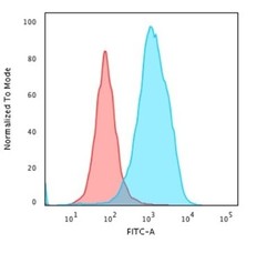
- Flow Cytometry: HSP60 Antibody (LK1) [NBP2-32973] - Flow Cytometric Analysis of paraformaldehyde-fixed HeLa cells using HSP60 Antibody (LK1) followed by goat anti-Mouse IgG-CF488 (Blue); Isotype Control (Red).
