Explore
 Validate
 Learn
Learn Western blot
 Immunocytochemistry
Antibody data
- Antibody Data
- Antigen structure
- References [3]
- Comments [0]
- Validations
- Western blot [1]
Submit
Validation data
Reference
Comment
Report error
- Product number
- M01280-3 - Provider product page

- Provider
- Boster Biological Technology
- Product name
- Anti-Hsp60/HSPD1 Antibody Picoband™ (monoclonal, 6G2)
- Antibody type
- Monoclonal
- Description
- Mouse IgG monoclonal antibody for Hsp60/HSPD1 detection. Tested with WB, IHC-P, ICC/IF, FCM in Human;Mouse;Rat.
- Reactivity
- Human, Mouse, Rat
- Host
- Mouse
- Isotype
- IgG
- Antibody clone number
- 6G2
- Vial size
- 100μg/vial
- Storage
- At -20°C for one year. After reconstitution, at 4°C for one month. It can also be aliquoted and stored frozen at -20°C for a longer time. Avoid repeated freezing and thawing.
- Handling
- Add 0.2ml of distilled water will yield a concentration of 500μg/ml.
Submitted references An experimental rat model of electric shock injury with isolated electric shock and water conduction: the histopathological changes on the skin and internal organs and the effect on biochemical parameters.
PRM-based quantitative proteomics analysis of altered HSP abundance in villi and decidua of patients with early missed abortion.
Molecular cloning, characterization, and expression of hsp60 in caudal fin regeneration of Misgurnus anguillicaudatus.
Dündar AS, Oruç M, Celbiş O, Şamdancı ET, Akatlı AN, Okumuş H, Taşkapan Ç, Özhan O, Parlakpınar H
International journal of legal medicine 2023 Jan;137(1):215-226
International journal of legal medicine 2023 Jan;137(1):215-226
PRM-based quantitative proteomics analysis of altered HSP abundance in villi and decidua of patients with early missed abortion.
Chen XF, Chen XQ, Luo HL, Xia LN, Huang SH, Chen Q
Proteome science 2023 Aug 16;21(1):12
Proteome science 2023 Aug 16;21(1):12
Molecular cloning, characterization, and expression of hsp60 in caudal fin regeneration of Misgurnus anguillicaudatus.
Li L, Nan P, Zhai S, Wang L, Si S, Chang Z
Molecular and cellular biochemistry 2014 Feb;387(1-2):143-50
Molecular and cellular biochemistry 2014 Feb;387(1-2):143-50
No comments: Submit comment
Supportive validation
- Submitted by
- Boster Biological Technology (provider)
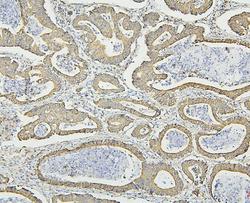
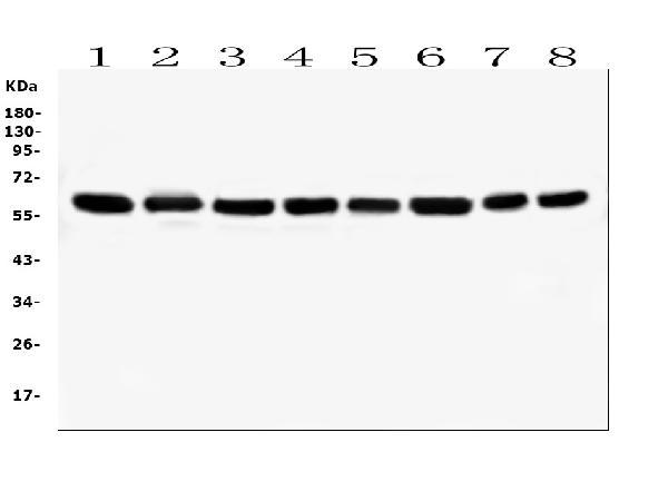
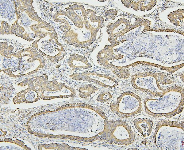
- Main image

- Experimental details
- Western blot analysis of HSPD1 using anti-HSPD1 antibody (M01280-3). Electrophoresis was performed on a 5-20% SDS-PAGE gel at 70V (Stacking gel) / 90V (Resolving gel) for 2-3 hours. The sample well of each lane was loaded with 50ug of sample under reducing conditions. Lane 1: human Caco-2 whole cell lysates Lane 2: human A549 whole cell lysates Lane 3: human THP-1 whole cell lysates Lane 4: human SW620 whole cell lysates Lane 5: human U-937 whole cell lysates Lane 6: human HepG2 whole cell lysates Lane 7: rat RH35 whole cell lysates Lane 8: mouse RAW246.7 whole cell lysates After Electrophoresis, proteins were transferred to a Nitrocellulose membrane at 150mA for 50-90 minutes. Blocked the membrane with 5% Non-fat Milk/ TBS for 1.5 hour at RT. The membrane was incubated with mouse anti-HSPD1 antigen affinity purified monoclonal antibody (Catalog # M01280-3) at 0.5 μg/mL overnight at 4°C, then washed with TBS-0.1%Tween 3 times with 5 minutes each and probed with a goat anti-mouse IgG-HRP secondary antibody at a dilution of 1:10000 for 1.5 hour at RT. The signal is developed using an Enhanced Chemiluminescent detection (ECL) kit (Catalog # EK1001) with Tanon 5200 system. A specific band was detected for HSPD1 at approximately 60KD. The expected band size for HSPD1 is at 60KD.
- Additional image