Explore
 Validate
 Learn
LearnMA3-012
antibody from Invitrogen Antibodies
Targeting: HSPD1
GroEL, HSP60, SPG13
 Western blot
 ELISA
ELISA Immunocytochemistry Immunoprecipitation Immunohistochemistry Flow cytometry Other assay
Antibody data
- Antibody Data
- Antigen structure
- References [19]
- Comments [0]
- Validations
- Immunocytochemistry [13]
- Immunoprecipitation [1]
- Immunohistochemistry [3]
- Flow cytometry [2]
- Other assay [2]
Submit
Validation data
Reference
Comment
Report error
- Product number
- MA3-012 - Provider product page

- Provider
- Invitrogen Antibodies
- Product name
- HSP60 Monoclonal Antibody (4B9/89)
- Antibody type
- Monoclonal
- Antigen
- Other
- Description
- MA3-012 detects heat shock protein 60 kDa (HSP60) from human, mouse, rat, and primate cells, tissues and recombinant protein preparations. MA3-012 has been successfully used in Western blot, flow cytometry, immunocytochemistry and ELISA procedures. By Western blot, this antibody detects a single 58 kDa protein representing HSP60 from human blood samples. Immunocytochemical staining of HSP60 in B-SC-1 cells with MA3-012 yields a pattern consistent with the mitochondrial localization of HSP60. The MA3-012 antigen is human placental HSP60. Epitope mapping studies using human HSP60 deletion mutants suggest that this antibody binds either between amino acids 335-366 or 484-547. Antibodies to this protein (and modification) were previously sold as part of a Thermo Scientific Cellomics High Content Screening Kit. This replacement antibody is now recommended for researchers who need an antibody for high content cell based assays. It has been thoroughly tested and validated for cellular immunofluorescence (IF) applications. Further optimization including the selection of the most appropriate fluorescent Dylight conjugated secondary antibody may have to be performed for your high content assay. Reconstitute in 100 µL PBS to create a stock of 1 mg/mL.
- Reactivity
- Human, Mouse, Rat
- Host
- Mouse
- Isotype
- IgG
- Antibody clone number
- 4B9/89
- Vial size
- 100 μg
- Concentration
- 1 mg/mL
- Storage
- -20°C, Avoid Freeze/Thaw Cycles
Submitted references UPR(mt) activation improves pathological alterations in cellular models of mitochondrial diseases.
The Role of Nonglycolytic Glucose Metabolism in Myocardial Recovery Upon Mechanical Unloading and Circulatory Support in Chronic Heart Failure.
A Study into the ADP-Ribosylome of IFN-γ-Stimulated THP-1 Human Macrophage-like Cells Identifies ARTD8/PARP14 and ARTD9/PARP9 ADP-Ribosylation.
Translation attenuation by minocycline enhances longevity and proteostasis in old post-stress-responsive organisms.
Multiplexed 3D super-resolution imaging of whole cells using spinning disk confocal microscopy and DNA-PAINT.
Fancd2 in vivo interaction network reveals a non-canonical role in mitochondrial function.
CCN6 regulates mitochondrial function.
Adipocytes from New Zealand obese mice exhibit aberrant proinflammatory reactivity to the stress signal heat shock protein 60.
Regulation of insulin secretion by SIRT4, a mitochondrial ADP-ribosyltransferase.
Synergistic and differential modulation of immune responses by Hsp60 and lipopolysaccharide.
The human silent information regulator (Sir)2 homologue hSIRT3 is a mitochondrial nicotinamide adenine dinucleotide-dependent deacetylase.
Differential expression of stress proteins in nonhuman primate lung and conducting airway after ozone exposure.
The identification of secreted heat shock 60 -like protein from rat glial cells and a human neuroblastoma cell line.
Simultaneous overexpression of two stress proteins in rat cardiomyocytes and myogenic cells confers protection against ischemia-induced injury.
Immunoelectron microscopic localization of the 60-kDa heat shock chaperonin protein (Hsp60) in mammalian cells.
Specific induction of the 70-kD heat stress proteins by the tyrosine kinase inhibitor herbimycin-A protects rat neonatal cardiomyocytes. A new pharmacological route to stress protein expression?
Chaperonin-mediated protein folding: GroES binds to one end of the GroEL cylinder, which accommodates the protein substrate within its central cavity.
Chaperonin-mediated protein folding: GroES binds to one end of the GroEL cylinder, which accommodates the protein substrate within its central cavity.
Primary structure of a human mitochondrial protein homologous to the bacterial and plant chaperonins and to the 65-kilodalton mycobacterial antigen.
Suárez-Rivero JM, Pastor-Maldonado CJ, Povea-Cabello S, Álvarez-Córdoba M, Villalón-García I, Talaverón-Rey M, Suárez-Carrillo A, Munuera-Cabeza M, Reche-López D, Cilleros-Holgado P, Piñero-Perez R, Sánchez-Alcázar JA
Orphanet journal of rare diseases 2022 May 17;17(1):204
Orphanet journal of rare diseases 2022 May 17;17(1):204
The Role of Nonglycolytic Glucose Metabolism in Myocardial Recovery Upon Mechanical Unloading and Circulatory Support in Chronic Heart Failure.
Badolia R, Ramadurai DKA, Abel ED, Ferrin P, Taleb I, Shankar TS, Krokidi AT, Navankasattusas S, McKellar SH, Yin M, Kfoury AG, Wever-Pinzon O, Fang JC, Selzman CH, Chaudhuri D, Rutter J, Drakos SG
Circulation 2020 Jul 21;142(3):259-274
Circulation 2020 Jul 21;142(3):259-274
A Study into the ADP-Ribosylome of IFN-γ-Stimulated THP-1 Human Macrophage-like Cells Identifies ARTD8/PARP14 and ARTD9/PARP9 ADP-Ribosylation.
Higashi H, Maejima T, Lee LH, Yamazaki Y, Hottiger MO, Singh SA, Aikawa M
Journal of proteome research 2019 Apr 5;18(4):1607-1622
Journal of proteome research 2019 Apr 5;18(4):1607-1622
Translation attenuation by minocycline enhances longevity and proteostasis in old post-stress-responsive organisms.
Solis GM, Kardakaris R, Valentine ER, Bar-Peled L, Chen AL, Blewett MM, McCormick MA, Williamson JR, Kennedy B, Cravatt BF, Petrascheck M
eLife 2018 Nov 27;7
eLife 2018 Nov 27;7
Multiplexed 3D super-resolution imaging of whole cells using spinning disk confocal microscopy and DNA-PAINT.
Schueder F, Lara-Gutiérrez J, Beliveau BJ, Saka SK, Sasaki HM, Woehrstein JB, Strauss MT, Grabmayr H, Yin P, Jungmann R
Nature communications 2017 Dec 12;8(1):2090
Nature communications 2017 Dec 12;8(1):2090
Fancd2 in vivo interaction network reveals a non-canonical role in mitochondrial function.
Zhang T, Du W, Wilson AF, Namekawa SH, Andreassen PR, Meetei AR, Pang Q
Scientific reports 2017 Apr 5;7:45626
Scientific reports 2017 Apr 5;7:45626
CCN6 regulates mitochondrial function.
Patra M, Mahata SK, Padhan DK, Sen M
Journal of cell science 2016 Jul 15;129(14):2841-51
Journal of cell science 2016 Jul 15;129(14):2841-51
Adipocytes from New Zealand obese mice exhibit aberrant proinflammatory reactivity to the stress signal heat shock protein 60.
Märker T, Kriebel J, Wohlrab U, Burkart V, Habich C
Journal of diabetes research 2014;2014:187153
Journal of diabetes research 2014;2014:187153
Regulation of insulin secretion by SIRT4, a mitochondrial ADP-ribosyltransferase.
Ahuja N, Schwer B, Carobbio S, Waltregny D, North BJ, Castronovo V, Maechler P, Verdin E
The Journal of biological chemistry 2007 Nov 16;282(46):33583-33592
The Journal of biological chemistry 2007 Nov 16;282(46):33583-33592
Synergistic and differential modulation of immune responses by Hsp60 and lipopolysaccharide.
Osterloh A, Kalinke U, Weiss S, Fleischer B, Breloer M
The Journal of biological chemistry 2007 Feb 16;282(7):4669-4680
The Journal of biological chemistry 2007 Feb 16;282(7):4669-4680
The human silent information regulator (Sir)2 homologue hSIRT3 is a mitochondrial nicotinamide adenine dinucleotide-dependent deacetylase.
Schwer B, North BJ, Frye RA, Ott M, Verdin E
The Journal of cell biology 2002 Aug 19;158(4):647-57
The Journal of cell biology 2002 Aug 19;158(4):647-57
Differential expression of stress proteins in nonhuman primate lung and conducting airway after ozone exposure.
Wu R, Zhao YH, Plopper CG, Chang MM, Chmiel K, Cross JJ, Weir A, Last JA, Tarkington B
The American journal of physiology 1999 Sep;277(3 Pt 1):L511-22
The American journal of physiology 1999 Sep;277(3 Pt 1):L511-22
The identification of secreted heat shock 60 -like protein from rat glial cells and a human neuroblastoma cell line.
Bassan M, Zamostiano R, Giladi E, Davidson A, Wollman Y, Pitman J, Hauser J, Brenneman DE, Gozes I
Neuroscience letters 1998 Jun 26;250(1):37-40
Neuroscience letters 1998 Jun 26;250(1):37-40
Simultaneous overexpression of two stress proteins in rat cardiomyocytes and myogenic cells confers protection against ischemia-induced injury.
Lau S, Patnaik N, Sayen MR, Mestril R
Circulation 1997 Oct 7;96(7):2287-94
Circulation 1997 Oct 7;96(7):2287-94
Immunoelectron microscopic localization of the 60-kDa heat shock chaperonin protein (Hsp60) in mammalian cells.
Soltys BJ, Gupta RS
Experimental cell research 1996 Jan 10;222(1):16-27
Experimental cell research 1996 Jan 10;222(1):16-27
Specific induction of the 70-kD heat stress proteins by the tyrosine kinase inhibitor herbimycin-A protects rat neonatal cardiomyocytes. A new pharmacological route to stress protein expression?
Morris SD, Cumming DV, Latchman DS, Yellon DM
The Journal of clinical investigation 1996 Feb 1;97(3):706-12
The Journal of clinical investigation 1996 Feb 1;97(3):706-12
Chaperonin-mediated protein folding: GroES binds to one end of the GroEL cylinder, which accommodates the protein substrate within its central cavity.
Langer T, Pfeifer G, Martin J, Baumeister W, Hartl FU
The EMBO journal 1992 Dec;11(13):4757-65
The EMBO journal 1992 Dec;11(13):4757-65
Chaperonin-mediated protein folding: GroES binds to one end of the GroEL cylinder, which accommodates the protein substrate within its central cavity.
Langer T, Pfeifer G, Martin J, Baumeister W, Hartl FU
The EMBO journal 1992 Dec;11(13):4757-65
The EMBO journal 1992 Dec;11(13):4757-65
Primary structure of a human mitochondrial protein homologous to the bacterial and plant chaperonins and to the 65-kilodalton mycobacterial antigen.
Jindal S, Dudani AK, Singh B, Harley CB, Gupta RS
Molecular and cellular biology 1989 May;9(5):2279-83
Molecular and cellular biology 1989 May;9(5):2279-83
No comments: Submit comment
Supportive validation
- Submitted by
- Invitrogen Antibodies (provider)
- Main image
- Experimental details
- Immunofluorescent analysis of Heat Shock Protein 60 using Anti-Heat Shock Protein 60 Monoclonal Antibody (4B9/89) (Product # MA3-012) shows staining in A2058 Cells. Heat Shock Protein 60 staining (green), F-Actin staining with Phalloidin (red) and nuclei with DAPI (blue) is shown. Cells were grown on chamber slides and fixed with formaldehyde prior to staining. Cells were probed without (control) or with or an antibody recognizing Heat Shock Protein 60 (Product # MA3-012) at a dilution of 1:200 over night at 4°C, washed with PBS and incubated with a DyLight-488 conjugated secondary antibody (Product # 35503, Goat Anti-Mouse). Images were taken at 60X magnification.
- Submitted by
- Invitrogen Antibodies (provider)
- Main image
- Experimental details
- Immunofluorescent analysis of Heat Shock Protein 60 using Anti-Heat Shock Protein 60 Monoclonal Antibody (4B9/89) (Product # MA3-012) shows staining in ATDC5 Cells. Heat Shock Protein 60 staining (green), F-Actin staining with Phalloidin (red) and nuclei with DAPI (blue) is shown. Cells were grown on chamber slides and fixed with formaldehyde prior to staining. Cells were probed without (control) or with or an antibody recognizing Heat Shock Protein 60 (Product # MA3-012) at a dilution of 1:100 over night at 4°C, washed with PBS and incubated with a DyLight-488 conjugated secondary antibody (Product # 35503, Goat Anti-Mouse). Images were taken at 60X magnification.

- Submitted by
- Invitrogen Antibodies (provider)
- Main image
- Experimental details
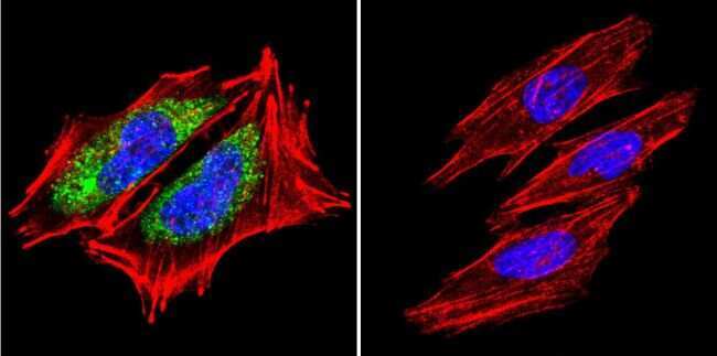
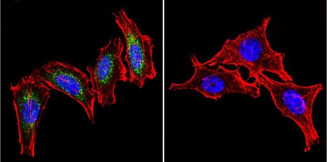
- Immunofluorescent analysis of Heat Shock Protein 60 using Anti-Heat Shock Protein 60 Monoclonal Antibody (4B9/89) (Product # MA3-012) shows staining in Hela Cells. Heat Shock Protein 60 staining (green), F-Actin staining with Phalloidin (red) and nuclei with DAPI (blue) is shown. Cells were grown on chamber slides and fixed with formaldehyde prior to staining. Cells were probed without (control) or with or an antibody recognizing Heat Shock Protein 60 (Product # MA3-012) at a dilution of 1:100 over night at 4°C, washed with PBS and incubated with a DyLight-488 conjugated secondary antibody (Product # 35503, Goat Anti-Mouse). Images were taken at 60X magnification.

- Submitted by
- Invitrogen Antibodies (provider)
- Main image

- Experimental details
- Immunofluorescent analysis of Heat Shock Protein 60 using a Hsp60 Monoclonal Antibody (Product # MA3-012) in human endothelial cells.
- Submitted by
- Invitrogen Antibodies (provider)
- Main image

- Experimental details
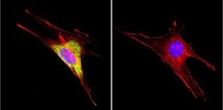
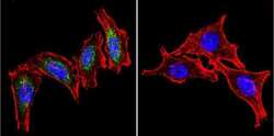
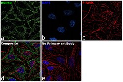
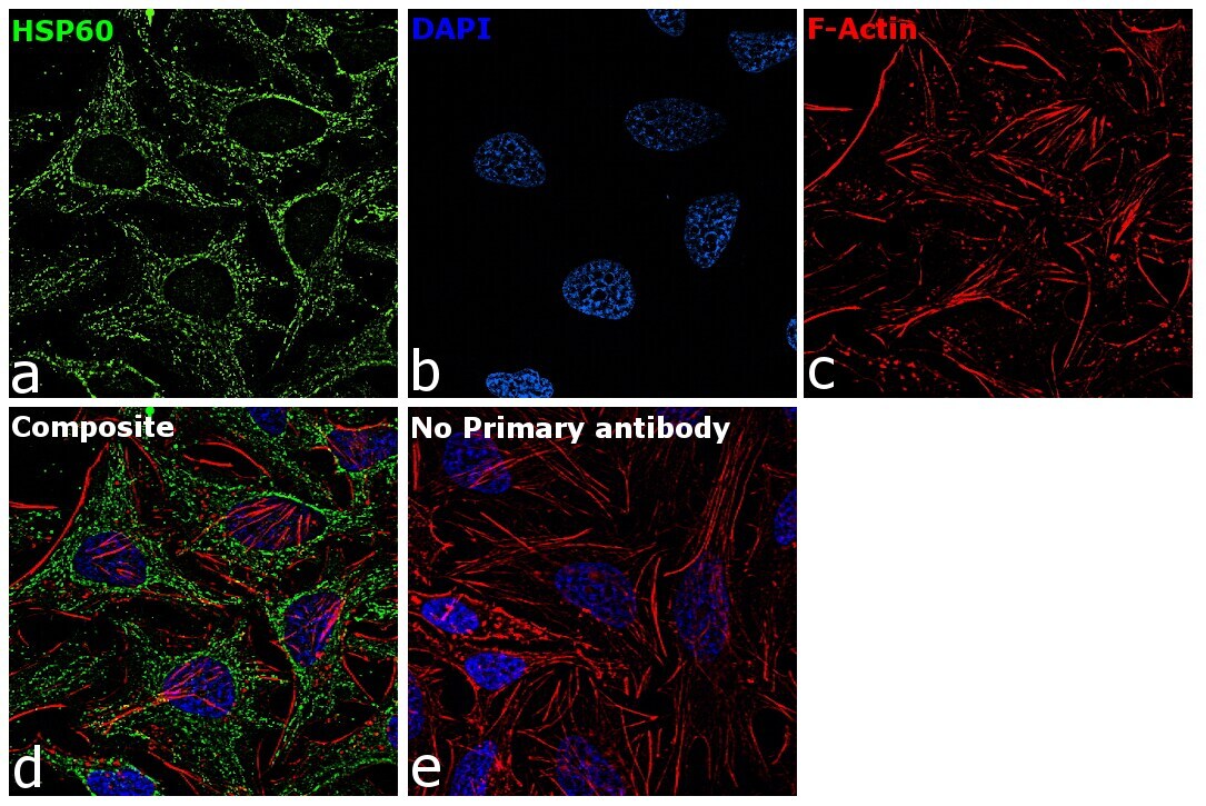
- Immunofluorescence analysis of HSP60 was performed using 70% confluent log phase HeLa cells. The cells were fixed with 4% paraformaldehyde for 10 minutes, permeabilized with 0.1% Triton™ X-100 for 15 minutes, and blocked with 2% BSA for 45 minutes at room temperature. The cells were labeled with HSP60 Monoclonal Antibody (4B9/89) (Product # MA3-012) at 1:60 dilution in 0.1% BSA, incubated at 4 degree celsius overnight and then labeled with Goat anti-Mouse IgG (H+L) Highly Cross-Adsorbed Secondary Antibody, Alexa Fluor Plus 488 (Product # A32723), (1:2000), for 45 minutes at room temperature (Panel a: Green). Nuclei (Panel b:Blue) were stained with ProLong™ Diamond Antifade Mountant with DAPI (Product # P36962). F-actin (Panel c: Red) was stained with Rhodamine Phalloidin (Product # R415, 1:300). Panel d represents the merged image showing cytoplasmic(mitochondrial like pattern) localization. Panel e represents control cells with no primary antibody to assess background. The images were captured at 60X magnification.

- Submitted by
- Invitrogen Antibodies (provider)
- Main image

- Experimental details
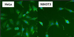
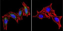
- Immunofluorescent analysis of Heat Shock Protein 60 (HSP60) (green) in HeLa and NIH3T3 cells. Formalin fixed cells were permeabilized with 0.1% Triton X-100 in TBS for 10 minutes at room temperature and blocked with 1% Blocker BSA (Product # 37525) for 15 minutes at room temperature. Cells were probed with a HSP60 monoclonal antibody (Product # MA3-012), at a dilution of 1:50 for at least 1 hour at room temperature, washed with PBS, and incubated with DyLight 488 goat-anti-mouse IgG secondary antibody (Product # 35502) at a dilution of 1:400 for 30 minutes at room temperature. Nuclei (blue) were stained with Hoechst 33342 dye (Product # 62249). Images were taken on a Thermo Scientific ArrayScan at 20X magnification.

- Submitted by
- Invitrogen Antibodies (provider)
- Main image

- Experimental details
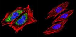
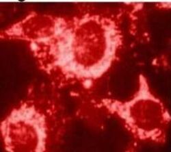
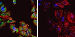
- Immunofluorescent analysis of Heat Shock Protein 60 (Hsp60, green) in HeLa cells. Formalin fixed cells were permeabilized with 0.1% Triton X-100 in TBS for 10 minutes at room temperature and blocked with 1% Blocker BSA (Product # 37525) for 15 minutes at room temperature. Cells were probed with (left panel) or without (right panel) a Hsp60 monoclonal antibody (Product # MA3-012), at a dilution of 1:50 for at least 1 hour at room temperature, washed with PBS, and incubated with DyLight 488 goat-anti-mouse IgG secondary antibody (Product # 35502) at a dilution of 1:400 for 30 minutes at room temperature. F-Actin (red) was stained with Dylight 554 phalloidin (Product # 21834), and nuclei (blue) were stained with Hoechst 33342 dye (Product # 62249). Images were taken on a Thermo Scientific ArrayScan or ToxInsight at 20X magnification.

- Submitted by
- Invitrogen Antibodies (provider)
- Main image
- Experimental details
- Immunofluorescent analysis of Heat Shock Protein 60 (HSP60) (green) in HeLa and NIH3T3 cells. Formalin fixed cells were permeabilized with 0.1% Triton X-100 in TBS for 10 minutes at room temperature and blocked with 1% Blocker BSA (Product # 37525) for 15 minutes at room temperature. Cells were probed with a HSP60 monoclonal antibody (Product # MA3-012), at a dilution of 1:50 for at least 1 hour at room temperature, washed with PBS, and incubated with DyLight 488 goat-anti-mouse IgG secondary antibody (Product # 35502) at a dilution of 1:400 for 30 minutes at room temperature. Nuclei (blue) were stained with Hoechst 33342 dye (Product # 62249). Images were taken on a Thermo Scientific ArrayScan at 20X magnification.

- Submitted by
- Invitrogen Antibodies (provider)
- Main image
- Experimental details
- Immunofluorescent analysis of Heat Shock Protein 60 using a Hsp60 Monoclonal Antibody (Product # MA3-012) in human endothelial cells.

- Submitted by
- Invitrogen Antibodies (provider)
- Main image
- Experimental details
- Immunofluorescent analysis of Heat Shock Protein 60 (Hsp60, green) in HeLa cells. Formalin fixed cells were permeabilized with 0.1% Triton X-100 in TBS for 10 minutes at room temperature and blocked with 1% Blocker BSA (Product # 37525) for 15 minutes at room temperature. Cells were probed with (left panel) or without (right panel) a Hsp60 monoclonal antibody (Product # MA3-012), at a dilution of 1:50 for at least 1 hour at room temperature, washed with PBS, and incubated with DyLight 488 goat-anti-mouse IgG secondary antibody (Product # 35502) at a dilution of 1:400 for 30 minutes at room temperature. F-Actin (red) was stained with Dylight 554 phalloidin (Product # 21834), and nuclei (blue) were stained with Hoechst 33342 dye (Product # 62249). Images were taken on a Thermo Scientific ArrayScan or ToxInsight at 20X magnification.

- Submitted by
- Invitrogen Antibodies (provider)
- Main image

- Experimental details
- Immunofluorescent analysis of Heat Shock Protein 60 using Anti-Heat Shock Protein 60 Monoclonal Antibody (4B9/89) (Product # MA3-012) shows staining in A2058 Cells. Heat Shock Protein 60 staining (green), F-Actin staining with Phalloidin (red) and nuclei with DAPI (blue) is shown. Cells were grown on chamber slides and fixed with formaldehyde prior to staining. Cells were probed without (control) or with or an antibody recognizing Heat Shock Protein 60 (Product # MA3-012) at a dilution of 1:200 over night at 4°C, washed with PBS and incubated with a DyLight-488 conjugated secondary antibody (Product # 35503, Goat Anti-Mouse). Images were taken at 60X magnification.

- Submitted by
- Invitrogen Antibodies (provider)
- Main image
- Experimental details
- Immunofluorescent analysis of Heat Shock Protein 60 using Anti-Heat Shock Protein 60 Monoclonal Antibody (4B9/89) (Product # MA3-012) shows staining in Hela Cells. Heat Shock Protein 60 staining (green), F-Actin staining with Phalloidin (red) and nuclei with DAPI (blue) is shown. Cells were grown on chamber slides and fixed with formaldehyde prior to staining. Cells were probed without (control) or with or an antibody recognizing Heat Shock Protein 60 (Product # MA3-012) at a dilution of 1:100 over night at 4°C, washed with PBS and incubated with a DyLight-488 conjugated secondary antibody (Product # 35503, Goat Anti-Mouse). Images were taken at 60X magnification.
- Submitted by
- Invitrogen Antibodies (provider)
- Main image
- Experimental details
- Immunofluorescence analysis of HSP60 was performed using 70% confluent log phase HeLa cells. The cells were fixed with 4% paraformaldehyde for 10 minutes, permeabilized with 0.1% Triton™ X-100 for 15 minutes, and blocked with 2% BSA for 45 minutes at room temperature. The cells were labeled with HSP60 Monoclonal Antibody (4B9/89) (Product # MA3-012) at 1:60 dilution in 0.1% BSA, incubated at 4 degree celsius overnight and then labeled with Goat anti-Mouse IgG (H+L) Highly Cross-Adsorbed Secondary Antibody, Alexa Fluor Plus 488 (Product # A32723), (1:2000), for 45 minutes at room temperature (Panel a: Green). Nuclei (Panel b:Blue) were stained with ProLong™ Diamond Antifade Mountant with DAPI (Product # P36962). F-actin (Panel c: Red) was stained with Rhodamine Phalloidin (Product # R415, 1:300). Panel d represents the merged image showing cytoplasmic(mitochondrial like pattern) localization. Panel e represents control cells with no primary antibody to assess background. The images were captured at 60X magnification.
Supportive validation
- Submitted by
- Invitrogen Antibodies (provider)
- Main image
- Experimental details
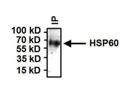
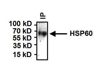
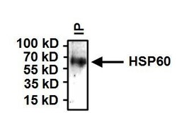
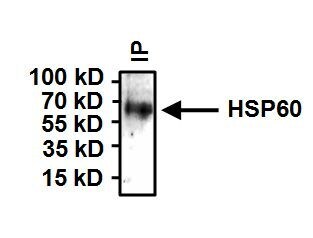
- Immunoprecipitation of Heat Shock Protein 60 (HSP60) was performed on HeLa cells. Antigen:antibody complexes were formed by incubating 500 µg whole cell lysate with 2 µg of HSP60 monoclonal antibody (Product # MA3-012) overnight on a rocking platform at 4°C. The immune complexes were captured on 50 µL Protein A/G Plus Agarose (Product # 20421), washed extensively, and eluted with Lane Marker Reducing Sample Buffer (Product # 39000). Samples were then resolved on a 4-20% Tris-HCl polyacrylamide gel, transferred to a PVDF membrane, and blocked with 5% BSA/TBST for at least 1 hour. The membrane was probed with a HSP60 monoclonal antibody (Product # MA3-012) at a dilution of 1:1000 overnight rotating at 4°C, washed in TBST, and probed with Clean-Blot IP Detection Reagent (HRP) at a dilution of 1:1000 (Product # 21230) for at least one hour. Chemiluminescent detection was performed using SuperSignal West Dura (Product # 34075).
Supportive validation
- Submitted by
- Invitrogen Antibodies (provider)
- Main image

- Experimental details
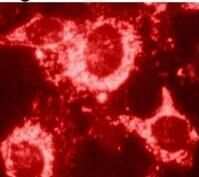
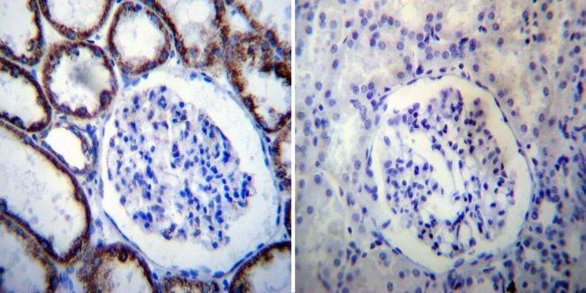
- Immunohistochemistry was performed on normal deparaffinized human Kidney tissue. To expose target proteins, heat induced antigen retrieval was performed using 10mM sodium citrate (pH6.0) buffer, microwaved for 8-15 minutes. Following antigen retrieval tissues were blocked in 3% BSA-PBS for 30 minutes at room temperature. Tissues were then probed at a dilution of 1:100 with a mouse monoclonal antibody recognizing Heat Shock Protein 60 (Product # MA3-012) or without primary antibody (negative control) overnight at 4°C in a humidified chamber. Tissues were washed extensively with PBST and endogenous peroxidase activity was quenched with a peroxidase suppressor. Detection was performed using a biotin-conjugated secondary antibody and SA-HRP, followed by colorimetric detection using DAB. Tissues were counterstained with hematoxylin and prepped for mounting.
- Submitted by
- Invitrogen Antibodies (provider)
- Main image
- Experimental details
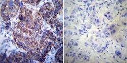
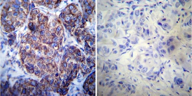
- Immunohistochemistry was performed on cancer biopsies of deparaffinized human Breast carcinoma tissue. To expose target proteins, heat induced antigen retrieval was performed using 10mM sodium citrate (pH6.0) buffer, microwaved for 8-15 minutes. Following antigen retrieval tissues were blocked in 3% BSA-PBS for 30 minutes at room temperature. Tissues were then probed at a dilution of 1:50 with a mouse monoclonal antibody recognizing Heat Shock Protein 60 (Product # MA3-012) or without primary antibody (negative control) overnight at 4°C in a humidified chamber. Tissues were washed extensively with PBST and endogenous peroxidase activity was quenched with a peroxidase suppressor. Detection was performed using a biotin-conjugated secondary antibody and SA-HRP, followed by colorimetric detection using DAB. Tissues were counterstained with hematoxylin and prepped for mounting.
- Submitted by
- Invitrogen Antibodies (provider)
- Main image
- Experimental details
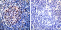
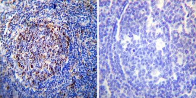
- Immunohistochemistry was performed on normal deparaffinized human Tonsil tissue. To expose target proteins, heat induced antigen retrieval was performed using 10mM sodium citrate (pH6.0) buffer, microwaved for 8-15 minutes. Following antigen retrieval tissues were blocked in 3% BSA-PBS for 30 minutes at room temperature. Tissues were then probed at a dilution of 1:100 with a mouse monoclonal antibody recognizing Heat Shock Protein 60 (Product # MA3-012) or without primary antibody (negative control) overnight at 4°C in a humidified chamber. Tissues were washed extensively with PBST and endogenous peroxidase activity was quenched with a peroxidase suppressor. Detection was performed using a biotin-conjugated secondary antibody and SA-HRP, followed by colorimetric detection using DAB. Tissues were counterstained with hematoxylin and prepped for mounting.
Supportive validation
- Submitted by
- Invitrogen Antibodies (provider)
- Main image
- Experimental details
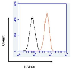
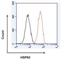
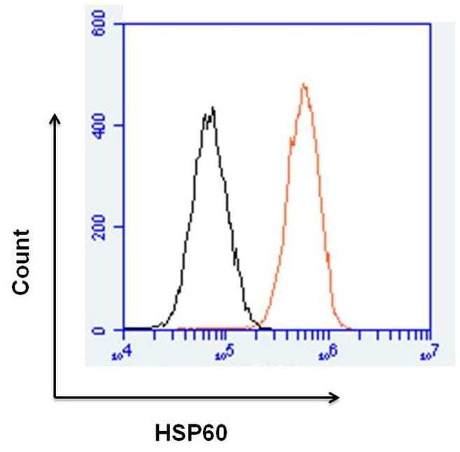
- Flow cytometry analysis of HSP60 was done on HeLa cells. Cells were fixed, permeabilized and stained with a HSP60 mouse monoclonal antibody (Product # MA3-012, orange histogram) or a mouse IgG2a isotype control (Product # MA1-10418, black histogram) at a dilution of 10 µg/mL. After incubation for 1 hour on ice, the cells were labeled with a Goat anti-Mouse IgG Secondary Antibody, DyLight 650 conjugate (Product # 84545) at a dilution of 1:50 for 1 hour on ice. A representative 10,000 cells were acquired and analyzed for each sample.

- Submitted by
- Invitrogen Antibodies (provider)
- Main image
- Experimental details
- Flow cytometry analysis of HSP60 was done on HeLa cells. Cells were fixed, permeabilized and stained with a HSP60 mouse monoclonal antibody (Product # MA3-012, orange histogram) or a mouse IgG2a isotype control (Product # MA1-10418, black histogram) at a dilution of 10 µg/mL. After incubation for 1 hour on ice, the cells were labeled with a Goat anti-Mouse IgG Secondary Antibody, DyLight 650 conjugate (Product # 84545) at a dilution of 1:50 for 1 hour on ice. A representative 10,000 cells were acquired and analyzed for each sample.
Supportive validation
- Submitted by
- Invitrogen Antibodies (provider)
- Main image
- Experimental details
- Immunoprecipitation of Heat Shock Protein 60 (HSP60) was performed on HeLa cells. Antigen:antibody complexes were formed by incubating 500 µg whole cell lysate with 2 µg of HSP60 monoclonal antibody (Product # MA3-012) overnight on a rocking platform at 4°C. The immune complexes were captured on 50 µL Protein A/G Plus Agarose (Product # 20421), washed extensively, and eluted with Lane Marker Reducing Sample Buffer (Product # 39000). Samples were then resolved on a 4-20% Tris-HCl polyacrylamide gel, transferred to a PVDF membrane, and blocked with 5% BSA/TBST for at least 1 hour. The membrane was probed with a HSP60 monoclonal antibody (Product # MA3-012) at a dilution of 1:1000 overnight rotating at 4°C, washed in TBST, and probed with Clean-Blot IP Detection Reagent (HRP) at a dilution of 1:1000 (Product # 21230) for at least one hour. Chemiluminescent detection was performed using SuperSignal West Dura (Product # 34075).
- Submitted by
- Invitrogen Antibodies (provider)
- Main image

- Experimental details
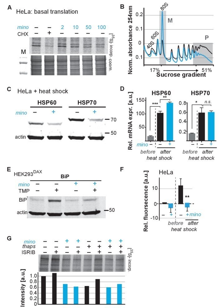
- Figure 4. Minocycline attenuates translation and reduces ribosomal load of highly translated mRNAs. ( A ) Autoradiograph (top) monitoring 35 S incorporation over 1 hr of HeLa cells treated for 6 hr with cycloheximide or increasing concentrations of minocycline. Coomassie gel (bottom) is shown as a loading control. Total of four independent experiments. ( B ) Polysome profile analysis of 12 hr water- (black) or minocycline-treated (blue) HeLa cells. High molecular weight polysomes ( P ) and 80S monosomes ( M ) regions are shaded in gray. An increase in monosomes and a decrease in high-molecular-weight polysomes are indicative of translation attenuation. Total of three independent experiments. ( C ) Immunoblots probing for HSP60 and HSP70 expression of water- or minocycline-treated (100 muM) HeLa cells after a 1 hr, 43degC heat shock. Total of three independent experiments. ( D ) qRT-PCR analysis of HSP60 and HSP70 mRNA expression in HeLa cells treated with or without minocycline (100 muM) before and after a 1 hr, 43degC heat shock. Minocycline treatment was initiated 12 hr prior to the heat shock. Data are represented as mean +- S.E.M. Significance determined by the Mann-Whitney t-test. Total of three independent experiments. ( E ) Immunoblots probing for BiP expression of water- or minocycline-treated (100 muM) HEK293 DAX cells engineered to allow UPR ER activation by trimethoprim treatment (TMP). Water or 100 muM minocycline was added alone or in combination with 20 uM t