Explore
 Validate
 Learn
Learn Western blot
Antibody data
- Antibody Data
- Antigen structure
- References [4]
- Comments [0]
- Validations
- Western blot [2]
- Immunocytochemistry [1]
Submit
Validation data
Reference
Comment
Report error
- Product number
- MAB1800 - Provider product page

- Provider
- R&D Systems
- Product name
- Human/Mouse/Rat HSP60 Antibody
- Antibody type
- Monoclonal
- Description
- Protein A or G purified from hybridoma culture supernatant. Detects human, mouse and rat HSP60.
- Reactivity
- Human, Mouse, Rat
- Host
- Mouse
- Conjugate
- Unconjugated
- Antigen sequence
P10809- Isotype
- IgG
- Antibody clone number
- 264233
- Vial size
- 100 ug
- Concentration
- LYOPH
- Storage
- Use a manual defrost freezer and avoid repeated freeze-thaw cycles. 12 months from date of receipt, -20 to -70 °C as supplied. 1 month, 2 to 8 °C under sterile conditions after reconstitution. 6 months, -20 to -70 °C under sterile conditions after reconstitution.
Submitted references MICU1 Confers Protection from MCU-Dependent Manganese Toxicity.
Comparative analysis of the interaction of HSPs in dendritic cells, macrophages, RGM-1 cells infected by Helicobacter pylori.
Heat shock protein 60: an endogenous inducer of dopaminergic cell death in Parkinson disease.
Nitric oxide-induced apoptosis in RAW 264.7 macrophages is mediated by endoplasmic reticulum stress pathway involving ATF6 and CHOP.
Wettmarshausen J, Goh V, Huang KT, Arduino DM, Tripathi U, Leimpek A, Cheng Y, Pittis AA, Gabaldón T, Mokranjac D, Hajnóczky G, Perocchi F
Cell reports 2018 Nov 6;25(6):1425-1435.e7
Cell reports 2018 Nov 6;25(6):1425-1435.e7
Comparative analysis of the interaction of HSPs in dendritic cells, macrophages, RGM-1 cells infected by Helicobacter pylori.
Yao Y, Wu J, Gu T, Cheng Y, Li G
American journal of translational research 2016;8(10):4184-4194
American journal of translational research 2016;8(10):4184-4194
Heat shock protein 60: an endogenous inducer of dopaminergic cell death in Parkinson disease.
Noelker C, Morel L, Osterloh A, Alvarez-Fischer D, Lescot T, Breloer M, Gold M, Oertel WH, Henze C, Michel PP, Dodel RC, Lu L, Hirsch EC, Hunot S, Hartmann A
Journal of neuroinflammation 2014 May 8;11:86
Journal of neuroinflammation 2014 May 8;11:86
Nitric oxide-induced apoptosis in RAW 264.7 macrophages is mediated by endoplasmic reticulum stress pathway involving ATF6 and CHOP.
Gotoh T, Oyadomari S, Mori K, Mori M
The Journal of biological chemistry 2002 Apr 5;277(14):12343-50
The Journal of biological chemistry 2002 Apr 5;277(14):12343-50
No comments: Submit comment
Supportive validation
- Submitted by
- R&D Systems (provider)
- Main image

- Experimental details
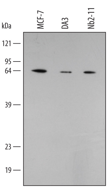
- Detection of Human/Mouse/Rat HSP60 by Western Blot. Western blot shows lysates of MCF-7 human breast cancer cell line, DA3 mouse myeloma cell line, and Nb2-11 rat lymphoma cell line. PVDF membrane was probed with 0.5 µg/mL of Mouse Anti-Human/Mouse/Rat HSP60 Monoclonal Antibody (Catalog # MAB1800) followed by HRP-conjugated Anti-Mouse IgG Secondary Antibody (Catalog # HAF007). A specific band was detected for HSP60 at approximately 62 kDa (as indicated). This experiment was conducted under reducing conditions and using Immunoblot Buffer Group 2.
- Submitted by
- R&D Systems (provider)
- Main image

- Experimental details
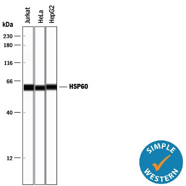
- Detection of Human HSP60 by Simple WesternTM. Simple Western lane view shows lysates of Jurkat human acute T cell leukemia cell line, HeLa human cervical epithelial carcinoma cell line, and HepG2 human hepatocellular carcinoma cell line, loaded at 0.2 mg/mL. A specific band was detected for HSP60 at approximately 60 kDa (as indicated) using 10 µg/mL of Mouse Anti-Human/Mouse/Rat HSP60 Monoclonal Antibody (Catalog # MAB1800). This experiment was conducted under reducing conditions and using the 12-230 kDa separation system.
Supportive validation
- Submitted by
- R&D Systems (provider)
- Main image

- Experimental details
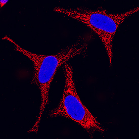
- HSP60 in HeLa Human Cell Line. HSP60 was detected in immersion fixed HeLa human cervical epithelial carcinoma cell line using Mouse Anti-Human/Mouse/Rat HSP60 Monoclonal Antibody (Catalog # MAB1800) at 25 µg/mL for 3 hours at room temperature. Cells were stained using the NorthernLights™ 557-conjugated Anti-Mouse IgG Secondary Antibody (red; Catalog # NL007) and counterstained with DAPI (blue). Specific staining was localized to cytoplasm. View our protocol for Fluorescent ICC Staining of Cells on Coverslips.