Explore
 Validate
 Learn
Learn Western blot
 Immunocytochemistry
Antibody data
- Antibody Data
- Antigen structure
- References [1]
- Comments [0]
- Validations
- Immunocytochemistry [3]
- Immunoprecipitation [1]
- Immunohistochemistry [1]
- Chromatin Immunoprecipitation [2]
- Other assay [3]
Submit
Validation data
Reference
Comment
Report error
- Product number
- PA5-28053 - Provider product page

- Provider
- Invitrogen Antibodies
- Product name
- ETS2 Polyclonal Antibody
- Antibody type
- Polyclonal
- Antigen
- Recombinant full-length protein
- Description
- Recommended positive controls: HeLa. Predicted reactivity: Mouse (87%), Rat (88%), Bovine (89%). Store product as a concentrated solution. Centrifuge briefly prior to opening the vial.
- Reactivity
- Human, Mouse, Rat
- Host
- Rabbit
- Isotype
- IgG
- Vial size
- 100 μL
- Concentration
- 0.35 mg/mL
- Storage
- Store at 4°C short term. For long term storage, store at -20°C, avoiding freeze/thaw cycles.
Submitted references ETS family transcriptional regulators drive chromatin dynamics and malignancy in squamous cell carcinomas.
Yang H, Schramek D, Adam RC, Keyes BE, Wang P, Zheng D, Fuchs E
eLife 2015 Nov 21;4:e10870
eLife 2015 Nov 21;4:e10870
No comments: Submit comment
Supportive validation
- Submitted by
- Invitrogen Antibodies (provider)
- Main image
- Experimental details
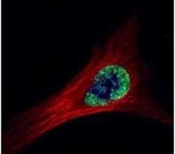
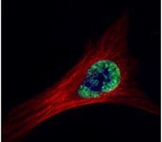
- Immunofluorescent analysis of ETS2 in paraformaldehyde-fixed HeLa cells using an ETS2 polyclonal antibody (Product # PA5-28053) (Green) at a 1:500 dilution. Alpha-tubulin filaments were labeled with Product # PA5-29281 (Red) at a 1:2000.
- Submitted by
- Invitrogen Antibodies (provider)
- Main image

- Experimental details
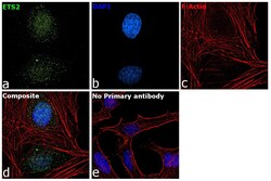
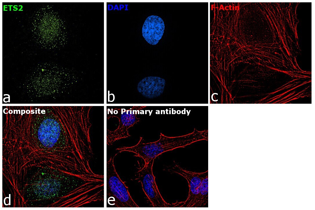
- Immunofluorescence analysis of ETS2 was performed using HeLa cells. The cells were fixed with 4% paraformaldehyde for 10 minutes, permeabilized with 0.1% Triton™ X-100 for 15 minutes, and blocked with 2% BSA for 1 hour at room temperature. The cells were labeled with ETS2 Polyclonal Antibody (Product # PA5-28053) at 1:200 dilution in 0.1% BSA and incubated overnight at 4 degree and then labeled with Goat anti-Rabbit IgG (H+L) Superclonal™ Recombinant Secondary Antibody, Alexa Fluor® 488 (Product # A27034, 1:2000 dilution) for 45 minutes at room temperature (Panel a: green) in HeLa cells. Nuclei (Panel b: blue) were stained with ProLong™ Diamond Antifade Mountant with DAPI (Product # P36962). F-actin (Panel c: red) was stained with Rhodamine Phalloidin (Product # R415, 1:300). Panel d represents the merged image of HeLa cells showing nuclear and cytoplasmic localization for ETS2. Panel e represents control cells with no primary antibody to assess background. The images were captured at 60X magnification.

- Submitted by
- Invitrogen Antibodies (provider)
- Main image
- Experimental details
- Immunofluorescence analysis of ETS2 was performed using HeLa cells. The cells were fixed with 4% paraformaldehyde for 10 minutes, permeabilized with 0.1% Triton™ X-100 for 15 minutes, and blocked with 2% BSA for 1 hour at room temperature. The cells were labeled with ETS2 Polyclonal Antibody (Product # PA5-28053) at 1:200 dilution in 0.1% BSA and incubated overnight at 4 degree and then labeled with Goat anti-Rabbit IgG (Heavy Chain) Superclonal™ Recombinant Secondary Antibody, Alexa Fluor® 488 (Product # A27034, 1:2000 dilution) for 45 minutes at room temperature (Panel a: green) in HeLa cells. Nuclei (Panel b: blue) were stained with ProLong™ Diamond Antifade Mountant with DAPI (Product # P36962). F-actin (Panel c: red) was stained with Rhodamine Phalloidin (Product # R415, 1:300). Panel d represents the merged image of HeLa cells showing nuclear and cytoplasmic localization for ETS2. Panel e represents control cells with no primary antibody to assess background. The images were captured at 60X magnification.
Supportive validation
- Submitted by
- Invitrogen Antibodies (provider)
- Main image
- Experimental details
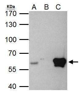
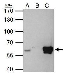
- ETS2 Polyclonal Antibody immunoprecipitates ETS2 protein in IP experiments. IP Sample: HeLa whole cell lysate/extract A. 40 µg HeLa whole cell lysate/extract B. Control with 2 µg of preimmune rabbit IgG C. Immunoprecipitation of ETS2 protein by 2 µg of ETS2 Polyclonal Antibody (Product # PA5-28053) 7.5% SDS-PAGE The immunoprecipitated ETS2 protein was detected by ETS2 Polyclonal Antibody (Product # PA5-28053) diluted at 1:1,000.

Supportive validation
- Submitted by
- Invitrogen Antibodies (provider)
- Main image
- Experimental details
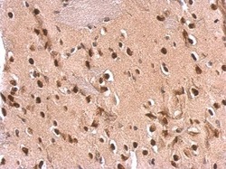
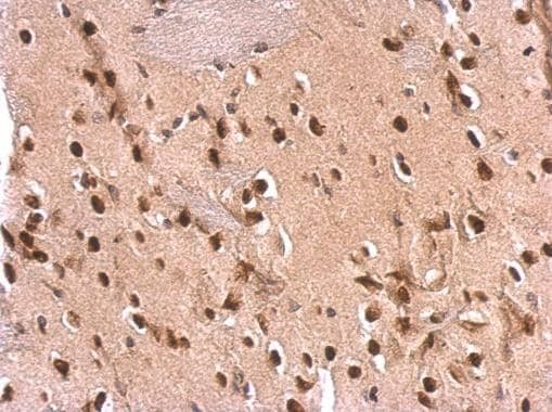
- ETS2 Polyclonal Antibody detects ETS2 protein at nucleus on rat fore brain by immunohistochemical analysis. Sample: Paraffin-embedded rat fore brain. ETS2 Polyclonal Antibody (Product # PA5-28053) dilution: 1:500. Antigen Retrieval: EDTA based buffer, pH 8.0, 15 min.
Supportive validation
- Submitted by
- Invitrogen Antibodies (provider)
- Main image
- Experimental details
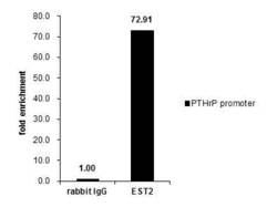
- Cross-linked ChIP was performed with MCF-7 chromatin extract and 5 µg of either control rabbit IgG or ETS2 Polyclonal Antibody (Product # PA5-28053). The precipitated DNA was detected by PCR with primer set targeting to PTHrP promoter.

- Submitted by
- Invitrogen Antibodies (provider)
- Main image

- Experimental details
- Cross-linked ChIP was performed with MCF-7 chromatin extract and 5 µg of either control rabbit IgG or ETS2 Polyclonal Antibody (Product # PA5-28053). The precipitated DNA was detected by PCR with primer set targeting to PTHrP promoter.

Supportive validation
- Submitted by
- Invitrogen Antibodies (provider)
- Main image

- Experimental details
- Figure 6. Super-activated ETS2 drives chromatin dynamics and transcriptional changes that occur during malignant transformation. ( A ) Summary of transcriptional profiling of basal epidermal progenitors (Epi-SC) purified from ETS2 (T72D) induced or control skin. Significantly upregulated genes (greater than twofold) were ranked and are listed at right with fold changes. Of note, SCC-SC super-enhancer (SE)-associated genes are marked in red. ( B ) Venn diagram showing significant overlap between differentially regulated transcripts in T72D-ETS2 Epi-SCs and SCC-SC as compared to Epi-SC. ( C ) Venn diagram showing that SEs of SCC-SCs show high overlap with those of ETS2 (T72D) Epi-SC. ( D ) Heatmap showing H3K27ac ChIP-seq read densities in the SCC-SC SEs. Note that read densities of ETS2 (T72D) induced Epi-SCs are higher than those of control Epi-SCs. ( E ) Examples of SEs acquired in ETS2 (T72D) induced Epi-SC and which show significant overlap with SCC-SC SEs. Shown are Elk3 and Mapk6 loci. ( F ) Examples of SEs shared not only by SCC-SCs and ETS2 (T72D)-EpiSCs, but also by wild-type EpiSCs. Note that both Neat1 and Cdh1 are highly expressed in both normal and malignant skin epithelia. ( G ) qPCR fold enrichment of ETS2 and myc ChIP DNA of SCC-SC super-enhancer epicenters. Values are normalized to IgG control (n = 3 +- SEM *p

- Submitted by
- Invitrogen Antibodies (provider)
- Main image

- Experimental details
- Figure 5--figure supplement 1. Validation of ETS2 expression constructs. ( A ) Lentiviral-based vector allowing for constitutive expression of a myc-tagged ETS2 cDNA, either WT or harboring silent mutations that abrogate shRNA binding. Immunoblot analysis to verify that both constructs express ETS2 protein at equivalent levels in HRas G12V ; Tgfbr2 - keratinocytes, but that when Ets2#649 shRNA is transduced, myc-ETS2 is only suppressed in cells transduced with the WT 3'UTR and not the mutant 3'UTR version. ( B ) Vector allowing for Doxycycline-inducible expression of a myc-tagged ETS2 cDNA, either WT or harboring the phosphomimetic T72D varient. H2B-RFP, driven by a constitutively active PGK promoter, was inserted to control for lentiviral transduction. When the vector is transduced in cells or mice expressing the rtTA Doxy-inducible transactivator, Doxy will activate the Myc-tagged, ETS2-T72D protein. Immunoblot analysis to verify ETS2 protein expression in HRas G12V ; Tgfbr2 - keratinocytes. ( C ) Ectopic induction of wild-type ETS2 in skin epidermis does not disrupt the morphology or function of the tissue. At E9.5, litters harboring K14rtTA and control embryos were infected in utero with lentivirus harboring the wild-type ETS2 construct in ( B ). This method allows for efficient and selective transduction of the skin epithelium by E12.5. Postnatally, these pups were administered Doxycycline to activate rtTA (if present) and 4 weeks later, their skin was

- Submitted by
- Invitrogen Antibodies (provider)
- Main image

- Experimental details
- ETS2 Polyclonal Antibody immunoprecipitates ETS2 protein in IP experiments. IP Sample: HeLa whole cell lysate/extract A. 40 µg HeLa whole cell lysate/extract B. Control with 2 µg of preimmune rabbit IgG C. Immunoprecipitation of ETS2 protein by 2 µg of ETS2 Polyclonal Antibody (Product # PA5-28053) 7.5% SDS-PAGE The immunoprecipitated ETS2 protein was detected by ETS2 Polyclonal Antibody (Product # PA5-28053) diluted at 1:1,000.