Explore
 Validate
 Learn
Learn Western blot
 ELISA
ELISAAntibody data
- Antibody Data
- Antigen structure
- References [0]
- Comments [0]
- Validations
- Western blot [2]
Submit
Validation data
Reference
Comment
Report error
- Product number
- PA1-21594 - Provider product page

- Provider
- Invitrogen Antibodies
- Product name
- Anti-Transferrin Polyclonal Antibody
- Antibody type
- Polyclonal
- Antigen
- Other
- Description
- PA1-21594 detects Transferrin in Human samples.
- Reactivity
- Human, Mouse, Bovine
- Host
- Rabbit
- Isotype
- IgG
- Vial size
- 100 µL
- Concentration
- 10 mg/mL
- Storage
- Store at 4°C short term. For long term storage, store at -20°C, avoiding freeze/thaw cycles.
No comments: Submit comment
Supportive validation
- Submitted by
- Invitrogen Antibodies (provider)
- Main image
- Experimental details
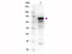
- Western blot detection of Transferrin in (lane 1) human Transferrin, (lane 2) control with no antibody using a Transferrin polyclonal antibody (Product # PA1-21594) at a dilution of 1:1000 overnight at 4°C. Secondary detection was performed using a Peroxidase rabbit secondary antibody at 1:40,000 for 30 min at room temperature and blocked for 30 min at room temperature. Predicted/Observed size: 76-81 kDa. 76 kDa for Transferrin. Other band (s): Transferrin slice varients and isoforms.
- Submitted by
- Invitrogen Antibodies (provider)
- Main image
- Experimental details
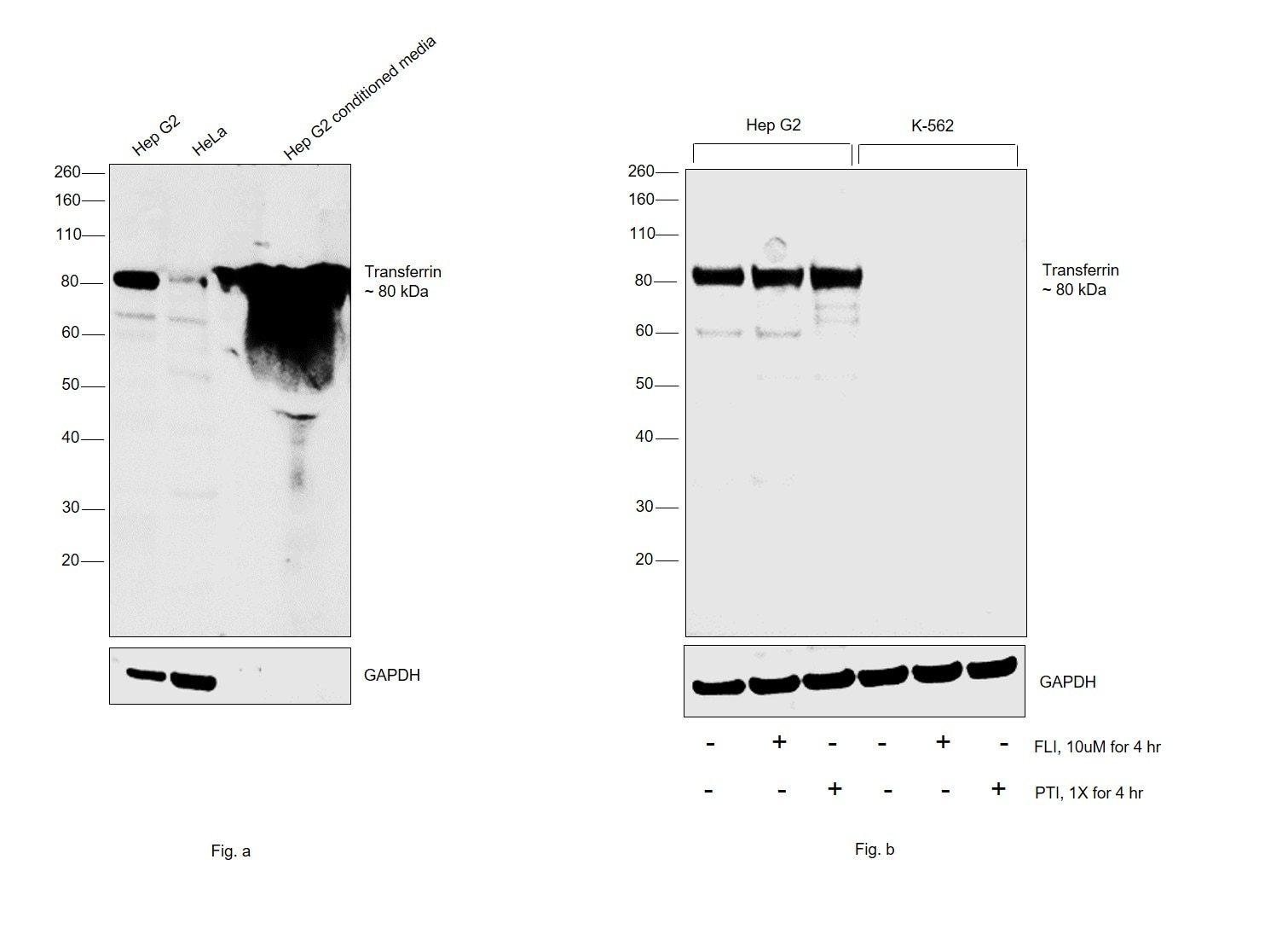
- Western blot was performed using Anti-Transferrin Polyclonal Antibody (Product # PA1-21594) and a 80 kDa band corresponding to Transferrin was observed across the samples in Fig. a and was also observed in Hep G2 cells upon treatment with secretion blockers (FLI-06 and PTI) in Fig. b. The expression of Transferrin was not observed in K-562 as reported in literature. An additional uncharacterized band was observed at ~60 kDa across all the samples tested. Whole cell extracts (30ug lysate) of Hep G2 (Lane 1), HeLa (Lane 2) and 7 ul of Hep G2 conditioned media (Lane 3) in Fig. a; whole cell extracts (30ug lysate) of Hep G2 (Lane 1) Hep G2 treated with FLI-06 (10uM for 4hr) (Lane 2), Hep G2 treated with PTI (1X for 4hr) (Lane 3), K-562 (Lane 4), K-562 treated with FLI-06 (10uM for 4hr) (Lane 5) and K-562 treated with PTI (1X for 4hr) (Lane 6) were electrophoresed using Novex® NuPAGE® 4-12% Bis-Tris Protein Gel (Product # NP0322BOX). Resolved proteins were then transferred onto a nitrocellulose membrane (Product # IB23001) by iBlot® 2 Dry Blotting System (Product # IB21001). The blot was probed with the primary antibody (1:500 dilution) and detected by chemiluminescence with Goat anti-Rabbit IgG (H+L), Superclonal™ Recombinant Secondary Antibody, HRP (Product # A27036, 1:4000 dilution) using the iBright FL 1000 (Product # A32752). Chemiluminescent detection was performed using Novex® ECL Chemiluminescent Substrate Reagent Kit (Product # WP20005).