 Explore
 Validate
 Learn
Learn Western blot
 ELISA
ELISAAntibody data
- Antibody Data
- Antigen structure
- References [0]
- Comments [0]
- Validations
- Western blot [3]
Submit
Validation data
Reference
Comment
Report error
- Product number
- LS-C745251 - Provider product page

- Provider
- LSBio
- Product name
- TF / Transferrin Antibody (Lyophilized, FITC) LS-C745251
- Antibody type
- Polyclonal
- Description
- Delipidation, salt fractionation and ion exchange chromatography followed by dialysis.
- Reactivity
- Human
- Host
- Rabbit
- Conjugate
- Green dye
- Isotype
- IgG
- Storage
- Store vial at -20°C or below prior to opening. Dilute 1:10 to minimize loss. Store the vial at -20°C or below after dilution. Avoid freeze-thaw cycles.
No comments: Submit comment
Enhanced validation
- Submitted by
- LSBio (provider)
- Enhanced method
- Genetic validation
- Main image

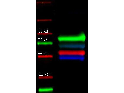
- Experimental details
- Rockland primary and Dylight conjugated secondary antibodies were used to detect: Human transferrin (1° 109-4134, green 2° 611-743-127); Alpha 1 anti trypsin (1° 100-101-147, red 2° 605-742-125); and Human IgG (1° 109-3102, Blue 2° 610-741-124 in a multiplex fluorescent western blot of human serum. Each primary antibody was diluted to 1:1000 in Blocking Buffer for Fluorescent Western Blotting - MB-070 and incubated for 2 hrs at RT. Blot was 3X in TTBS, 1X in TBS and probed with secondary antibodies diluted 1:10000) in MB-070 and incubated ~ 1hr at 4 degrees. After wash 2X in TTBS and 2X in TBS, blot was rinsed 2X in MeOH, dried and imaged using the Biorad VersaDoc4000.
- Submitted by
- LSBio (provider)
- Enhanced method
- Genetic validation
- Main image

- Experimental details
- Rabbit anti-Transferrin (109-4134 lot 3033, green), Goat-anti-Alpha-1-Anti-Trypsin (100-101-147 lot 5842), and Mouse-a-GST (200-301-200 lot 24882) were used in a multiplex system to detect target proteins under reducing (R) conditions (+4% BME) in albumin depleted human serum with 320 ng of added GST. Sample was run by SDS-PAGE, transferred to 0.2 um PVDF using the BioRad Trans-Blot Turbo and blocked in 2.5% Blotto, 2.5% BSA, 0.02% Tween over night at 4°C. Membrane was probed with three primary antibodies at 1:1000 dilution (in MB-070 over night at 4°C). Detection shown was using DyLight549 Donkey anti-Rabbit IgG (611-742-127 lot 21100, shown as green) DyLight 488 Donkey anti-Mouse IgG (610-741-124 lot 21095, shown as blue), and DyLight 649 Donkey anti-Goat IgG (605-743-125 lot 20834, shown as red) at 1:10000 (in MB-070 30 min RT). Blots were washed, rinsed in methanol, dried and Images were collected using the BioRad VersaDoc System.

- Submitted by
- LSBio (provider)
- Enhanced method
- Genetic validation
- Main image

- Experimental details
- Rockland primary and Dylight conjugated secondary antibodies were used to detect: Human transferrin (1° 109-4134, green 2° 611-743-127); Alpha 1 anti trypsin (1° 100-101-147, red 2° 605-742-125); and Human IgG (1° 109-3102, Blue 2° 610-741-124 in a multiplex fluorescent western blot of human serum. Each primary antibody was diluted to 1:1000 in Blocking Buffer for Fluorescent Western Blotting - MB-070 and incubated for 2 hrs at RT. Blot was 3X in TTBS, 1X in TBS and probed with secondary antibodies diluted 1:10000) in MB-070 and incubated ~ 1hr at 4 degrees. After wash 2X in TTBS and 2X in TBS, blot was rinsed 2X in MeOH, dried and imaged using the Biorad VersaDoc4000.
