Explore
 Validate
 Learn
Learn Western blot
 ELISA
ELISA Immunohistochemistry
Antibody data
- Antibody Data
- Antigen structure
- References [0]
- Comments [0]
- Validations
- Western blot [3]
- Immunohistochemistry [1]
Submit
Validation data
Reference
Comment
Report error
- Product number
- LS-C745249 - Provider product page

- Provider
- LSBio
- Product name
- TF / Transferrin Antibody (Lyophilized) LS-C745249
- Antibody type
- Polyclonal
- Description
- Delipidation, salt fractionation and ion exchange chromatography followed by dialysis.
- Reactivity
- Human
- Host
- Rabbit
- Isotype
- IgG
- Storage
- Store vial at -20°C or below prior to opening. Dilute 1:10 to minimize loss. Store the vial at -20°C or below after dilution. Avoid freeze-thaw cycles.
No comments: Submit comment
Enhanced validation
- Submitted by
- LSBio (provider)
- Enhanced method
- Genetic validation
- Main image
- Experimental details
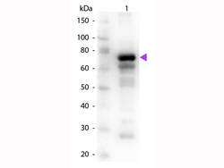
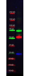
- Western Blot of rabbit Anti-Transferrin primary antibody. Lane 1: Human Transferrin. Lane 2: None. Primary antibody: Transferrin primary antibody at 1:1,000 overnight at 4°C. Secondary antibody: Peroxidase rabbit secondary antibody at 1:40,000 for 30 min at RT. Blocking: MB-070 for 30 min at RT. Predicted/Observed size: 76-81 kDa. 76 kDa for Transferrin. Other band(s): Transferrin slice varients and isoforms.
- Submitted by
- LSBio (provider)
- Enhanced method
- Genetic validation
- Main image
- Experimental details
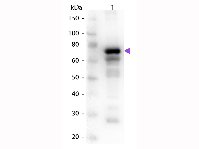
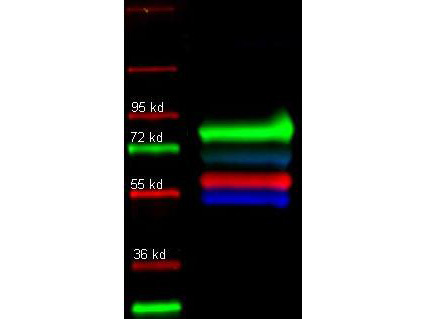
- Western Blot of rabbit anti-Transferrin antibody. Lane 1: molecular weight. Lane 2: anti-Transferrin (green), Goat-anti-Alpha-1-Anti-Trypsin, and Mouse-a-GST were used in a multiplex system to detect target proteins under reducing conditions in albumin depleted human serum with 320 ng of added GST. Load: 35 µg per lane. Primary antibody: Transferrin antibody at 1:1000 for overnight at 4°C. Secondary antibody: DyLight549 Donkey anti-Rabbit IgG (green), DyLight 488 Donkey anti-Mouse IgG (blue), and DyLight 649 Donkey anti-Goat IgG (red) at 1:10,000 for 45 min at RT. Block: 2.5% Blotto, 2.5% BSA, 0.02% Tween over night at 4°C. Other band(s): none.
- Submitted by
- LSBio (provider)
- Enhanced method
- Genetic validation
- Main image

- Experimental details
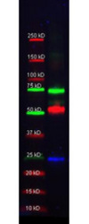
- Western Blot of primary and Dylight conjugated secondary antibodies. Lane 1: molecular weight. Lane 2: Rabbit anti-Human transferrin (green); Alpha 1 anti trypsin (red); and Human IgG (Blue) in a multiplex fluorescent western blot of human serum. Primary antibody: Each primary antibody was diluted to 1:1000 for 2 hrs at RT. Secondary antibody: Rabbit IgG (H&L) Antibody DyLight 649 Conjugated (green), Goat IgG (H&L) Antibody DyLight 549 Conjugated (red), Mouse IgG (H&L) Antibody DyLight 488 Conjugated (blue)secondary antibody at 1:10,000 for 45 min at RT. Block: IRdye blocking buffer (MB-070) 1hr at 4°C. Other band(s): none.
Supportive validation
- Submitted by
- LSBio (provider)
- Main image
- Experimental details
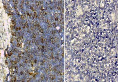
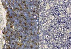
- Immunohistochemistry of rabbit anti human Transferrin antibody. Tissue: Proliferant, cycling human thymus. Antigen Retrieval: HIER pH6.2. Staining: diffuse cytoplasmic and membrane staining. Right: Transferrin antibody 209-4134 at 1:100. Left: Normal Rabbit IgG Negative Control.