Explore
 Validate
 Learn
Learn Western blot
Antibody data
- Antibody Data
- Antigen structure
- References [1]
- Comments [0]
- Validations
- Western blot [3]
- Immunocytochemistry [3]
Submit
Validation data
Reference
Comment
Report error
- Product number
- PA3-913 - Provider product page

- Provider
- Invitrogen Antibodies
- Product name
- Anti-Transferrin Polyclonal Antibody
- Antibody type
- Polyclonal
- Antigen
- Other
- Description
- PA3-913 detects transferrin from human and mouse samples. PA3-913 has been successfully used in Western Blot and ICC/IF procedures. The PA3-913 immunogen is human transferrin isolated from plasma.
- Reactivity
- Human, Mouse
- Host
- Rabbit
- Isotype
- IgG
- Vial size
- 100 µL
- Concentration
- Conc. Not Determined
- Storage
- -20° C, Avoid Freeze/Thaw Cycles
Submitted references Neuropathology of sporadic Parkinson disease before the appearance of parkinsonism: preclinical Parkinson disease.
Ferrer I, Martinez A, Blanco R, Dalfó E, Carmona M
Journal of neural transmission (Vienna, Austria : 1996) 2011 May;118(5):821-39
Journal of neural transmission (Vienna, Austria : 1996) 2011 May;118(5):821-39
No comments: Submit comment
Supportive validation
- Submitted by
- Invitrogen Antibodies (provider)
- Main image
- Experimental details
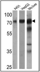
- Western blot analysis of Transferrin was performed by loading 25 µg of A431 (Lane 1), HepG2 (Lane 2) and mouse liver (Lane 3) cell lysates onto an SDS polyacrylamide gel. Proteins were transferred to a PVDF membrane and blocked at 4ºC overnight. The membrane was probed with a Transferrin polyclonal antibody (Product # PA3-913) at a dilution of 1:1000 (A431) and 1:2000 (HepG2 and mouse) overnight at 4°C, washed in TBST, and probed with an HRP-conjugated secondary antibody for 1 hr at room temperature in the dark. Chemiluminescent detection was performed using Pierce ECL Plus Western Blotting Substrate (Product # 32132). Results show a band at approx. 77 kDa.

- Submitted by
- Invitrogen Antibodies (provider)
- Main image

- Experimental details
- Western blot was performed using Anti-Transferrin Polyclonal Antibody (Product # PA3-913) and an 80 kDa band corresponding to Transferrin was observed in HepG2 cells upon treatment with secretion blockers (FLI-06 and PTI). The expression of Transferrin was not observed in K-562 which is a negative model as reported. An uncharacterized band at ~90kDa was observed in all the samples tested. Whole cell extracts (30ug lysate) of Hep G2 (Lane 1) Hep G2 treated with FLI-06 (10uM for 4hr) (Lane 2), Hep G2 treated with PTI (1X for 4hr) (Lane 3), K-562 (Lane 4), K-562 treated with FLI-06 (10uM for 4hr) (Lane 5) and K-562 treated with PTI (1X for 4hr) (Lane 6) were electrophoresed using Novex® NuPAGE® 4-12% Bis-Tris Protein Gel (Product # NP0322BOX). Resolved proteins were then transferred onto a nitrocellulose membrane (Product # IB23001) by iBlot® 2 Dry Blotting System (Product # IB21001). The blot was probed with the primary antibody (1:1000 dilution) and detected by chemiluminescence with Goat anti-Rabbit IgG (H+L) Superclonal™ Recombinant Secondary Antibody, HRP (Product # A27036, 1:4000 dilution) using the iBright FL 1000 (Product # A32752). Chemiluminescent detection was performed using Novex® ECL Chemiluminescent Substrate Reagent Kit (Product # WP20005).

- Submitted by
- Invitrogen Antibodies (provider)
- Main image

- Experimental details
- Western blot analysis was performed on Conditioned Medium-HeLa (Lane 1), Conditioned Medium-HepG2 (Lane 2) and Human Plasma (Lane 3). The blot was probed with Anti-Transferrin Polyclonal Antibody (Product # PA3-913, 1:1000 dilution) and detected by chemiluminescence using Goat anti-Rabbit IgG (H+L) Superclonal™ Secondary Antibody, HRP conjugate (Product # A27036, 0.25 µg/mL, 1:4000 dilution). A 70kDa band corresponding to Transferrin was seen in both conditioned medium tested as well as in Human Plasma.

Supportive validation
- Submitted by
- Invitrogen Antibodies (provider)
- Main image
- Experimental details
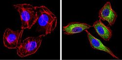
- Immunofluorescent analysis of Transferrin (green) showing staining in the secretion of U251 cells. Formalin-fixed cells were permeabilized with 0.1% Triton X-100 in TBS for 5-10 minutes and blocked with 3% BSA-PBS for 30 minutes at room temperature. Cells were probed with a Transferrin polyclonal antibody (Product # PA3-913) in 3% BSA-PBS at a dilution of 1:100 and incubated overnight at 4 ºC in a humidified chamber. Cells were washed with PBST and incubated with a DyLight-conjugated secondary antibody in PBS at room temperature in the dark. F-actin (red) was stained with a fluorescent red phalloidin and nuclei (blue) were stained with Hoechst or DAPI. Images were taken at a magnification of 60x.

- Submitted by
- Invitrogen Antibodies (provider)
- Main image

- Experimental details
- Immunofluorescent analysis of Transferrin (green) showing staining in the secretion of NIH-3T3 cells. Formalin-fixed cells were permeabilized with 0.1% Triton X-100 in TBS for 5-10 minutes and blocked with 3% BSA-PBS for 30 minutes at room temperature. Cells were probed with a Transferrin polyclonal antibody (Product # PA3-913) in 3% BSA-PBS at a dilution of 1:100 and incubated overnight at 4 ºC in a humidified chamber. Cells were washed with PBST and incubated with a DyLight-conjugated secondary antibody in PBS at room temperature in the dark. F-actin (red) was stained with a fluorescent red phalloidin and nuclei (blue) were stained with Hoechst or DAPI. Images were taken at a magnification of 60x.

- Submitted by
- Invitrogen Antibodies (provider)
- Main image

- Experimental details
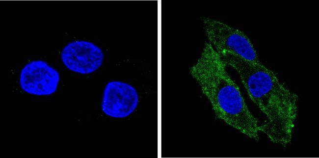
- Immunofluorescent analysis of Transferrin (green) showing staining in the secretion of HepG2 cells. Formalin-fixed cells were permeabilized with 0.1% Triton X-100 in TBS for 5-10 minutes and blocked with 3% BSA-PBS for 30 minutes at room temperature. Cells were probed with a Transferrin polyclonal antibody (Product # PA3-913) in 3% BSA-PBS at a dilution of 1:100 and incubated overnight at 4 ºC in a humidified chamber. Cells were washed with PBST and incubated with a DyLight-conjugated secondary antibody in PBS at room temperature in the dark. F-actin (red) was stained with a fluorescent red phalloidin and nuclei (blue) were stained with Hoechst or DAPI. Images were taken at a magnification of 60x.