Antibody data
- Antibody Data
- Antigen structure
- References [1]
- Comments [0]
- Validations
- Western blot [4]
- Immunohistochemistry [1]
- Other assay [1]
Submit
Validation data
Reference
Comment
Report error
- Product number
- PA5-27306 - Provider product page
- Provider
- Invitrogen Antibodies
- Product name
- Transferrin Polyclonal Antibody
- Antibody type
- Polyclonal
- Antigen
- Recombinant protein fragment
- Description
- Recommended positive controls: 293T, A431, H1299, HeLa, HepG2, Molt-4, Raji, human plasma, mouse plasma. Predicted reactivity: Chimpanzee (98%). Store product as a concentrated solution. Centrifuge briefly prior to opening the vial.
- Reactivity
- Human, Mouse
- Host
- Rabbit
- Isotype
- IgG
- Vial size
- 100 µL
- Concentration
- 0.12 mg/mL
- Storage
- Store at 4°C short term. For long term storage, store at -20°C, avoiding freeze/thaw cycles.
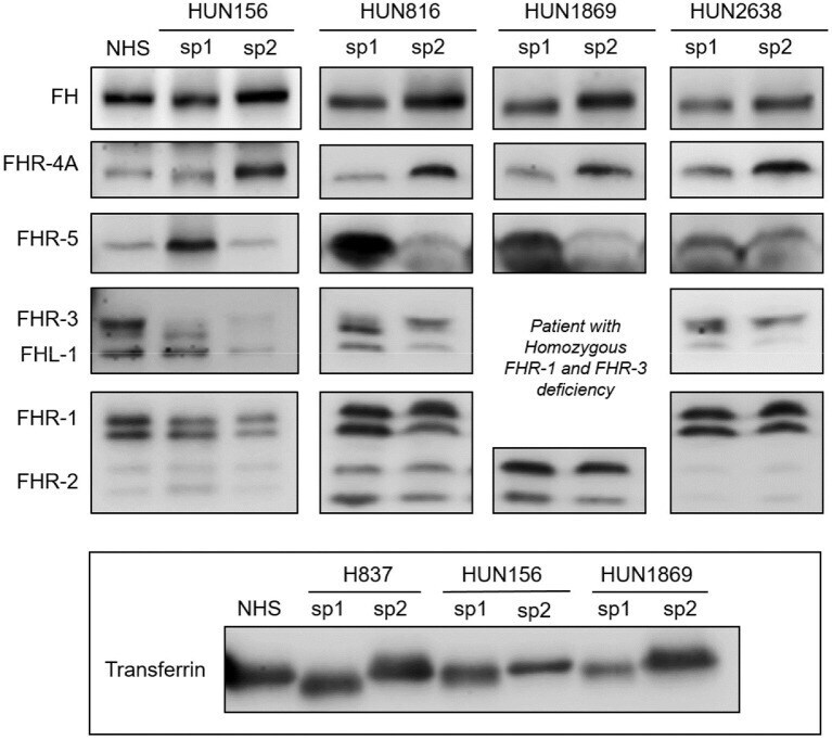
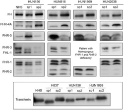
Submitted references Complement Genetic Variants and FH Desialylation in S. pneumoniae-Haemolytic Uraemic Syndrome.
Gómez Delgado I, Corvillo F, Nozal P, Arjona E, Madrid Á, Melgosa M, Bravo J, Szilágyi Á, Csuka D, Veszeli N, Prohászka Z, Sánchez-Corral P
Frontiers in immunology 2021;12:641656
No comments: Submit comment
Supportive validation
- Submitted by
- Invitrogen Antibodies (provider)
- Main image
- Experimental details
- Western Blot analysis of Transferrin was performed by separating 50 µg of Mouse tissue extracts by 7.5% SDS-PAGE. Proteins were transferred to a membrane and probed with a Transferrin Polyclonal Antibody (Product # PA5-27306) at a dilution of 1:1000. The HRP-conjugated anti-rabbit IgG antibody was used to detect the primary antibody.
- Submitted by
- Invitrogen Antibodies (provider)
- Main image
- Experimental details
- Western Blot using Transferrin Polyclonal Antibody (Product # PA5-27306). Sample (30 µg of whole cell lysate). Lane A: A431. Lane B: H1299. Lane C: HeLa. 7.5% SDS PAGE. Transferrin Polyclonal Antibody (Product # PA5-27306) diluted at 1:1,000. The HRP-conjugated anti-rabbit IgG antibody was used to detect the primary antibody.
- Submitted by
- Invitrogen Antibodies (provider)
- Main image
- Experimental details
- Western Blot analysis of Transferrin was performed by separating 30 µg of Human tissue extracts by 7.5% SDS-PAGE. Proteins were transferred to a membrane and probed with a Transferrin Polyclonal Antibody (Product # PA5-27306) at a dilution of 1:5000. The HRP-conjugated anti-rabbit IgG antibody was used to detect the primary antibody.
- Submitted by
- Invitrogen Antibodies (provider)
- Main image
- Experimental details
- Western blot was performed using Anti-Transferrin Polyclonal Antibody (Product # PA5-27306) and a 80 kDa band corresponding to Transferrin was observed across the samples except in HeLa and HeLa conditioned media in Fig. a and was also observed in Hep G2 cells upon treatment with secretion blockers (FLI-06 and PTI) in Fig. b. The expression of Transferrin was not observed in K-562 as reported in literature. Whole cell extracts of Hep G2 (Lane 1), HeLa (Lane 2), 7 ul of Hep G2 conditioned media (Lane 3), 7 ul of HeLa conditioned media (Lane 4), Mouse Liver (Lane 5), Rat Liver (Lane 6) and 2ul of Human Plasma (Lane 7) in Fig. a; whole cell extracts (30ug lysate) of Hep G2 (Lane 1) Hep G2 treated with FLI-06 (10uM for 4hr) (Lane 2), Hep G2 treated with PTI (1X for 4hr) (Lane 3), K-562 (Lane 4), K-562 treated with FLI-06 (10uM for 4hr) (Lane 5) and K-562 treated with PTI (1X for 4hr) (Lane 6) were electrophoresed using Novex® NuPAGE® 4-12% Bis-Tris Protein Gel (Product # NP0322BOX). Resolved proteins were then transferred onto a nitrocellulose membrane (Product # IB23001) by iBlot® 2 Dry Blotting System (Product # IB21001). The blot was probed with the primary antibody (1:1000 dilution) and detected by chemiluminescence with Goat anti-Rabbit IgG (H+L), Superclonal™ Recombinant Secondary Antibody, HRP (Product # A27036, 1:4000 dilution) using the iBright FL 1000 (Product # A32752). Chemiluminescent detection with Novex® ECL Kit (Product # WP20005).
Supportive validation
- Submitted by
- Invitrogen Antibodies (provider)
- Main image
- Experimental details
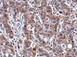
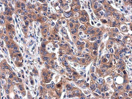
- Transferrin Polyclonal Antibody detects Transferrin protein at cytoplasm in human esophageal carcinoma by immunohistochemical analysis. Sample: Paraffin-embedded human esophageal carcinoma. Transferrin Polyclonal Antibody (Product # PA5-27306) diluted at 1:500. Antigen Retrieval: Citrate buffer, pH 6.0, 15 min.
Supportive validation
- Submitted by
- Invitrogen Antibodies (provider)
- Main image
- Experimental details
- Figure 3 Desialylated FH/FHRs in samples from Hungarian SP-HUS patients. WB analyses of FH/FHRs in two plasma samples (sp1 and sp2) from four Hungarian patients. Time between disease onset and extraction date was as follows: HUN156 (10 days and 11 months); HUN816 (9 days and 2 months); HUN1869 (5 days and 2 months), this patient is homozygous for FHR-3 and FHR-1 deficiency; HUN2638 (23 days and 43 days). WB analysis of human Transferrin (Mw 77 kDa) in sp1 and sp2 samples from patients H837, HUN156, and HUN 1869 is shown at the bottom inset.