 Explore
 Validate
 Learn
Learn Western blot
 ELISA
ELISA Immunocytochemistry
Antibody data
- Antibody Data
- Antigen structure
- References [0]
- Comments [0]
- Validations
- Immunocytochemistry [4]
- Flow cytometry [2]
Submit
Validation data
Reference
Comment
Report error
- Product number
- MA5-29035 - Provider product page

- Provider
- Invitrogen Antibodies
- Product name
- Apolipoprotein H Monoclonal Antibody (4A2F8H12)
- Antibody type
- Monoclonal
- Antigen
- Recombinant full-length protein
- Description
- This product is preservative free. It is recommended to add sodium azide to avoid contamination (final concentration 0.05%-0.1%). This antibody has specificity for Human APOH/B2G1.
- Reactivity
- Human
- Host
- Mouse
- Isotype
- IgG
- Antibody clone number
- 4A2F8H12
- Vial size
- 100 μL
- Concentration
- 1 mg/mL
- Storage
- Store at 4°C short term. For long term storage, store at -20°C, avoiding freeze/thaw cycles.
No comments: Submit comment
Supportive validation
- Submitted by
- Invitrogen Antibodies (provider)
- Main image
- Experimental details
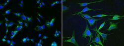
- Immunofluorescence staining of Human Apolipoprotein H in HepG2 or Hela cells. Cells were fixed with 4% PFA, permeabilzed with 1% Triton X-100 in PBS, blocked with 10% serum, and incubated with Apolipoprotein H Monoclonal Antibody (4A2F8H12) (Product # MA5-29035, 1:60). Then cells were stained with the Alexa Fluor® 488-conjugated Goat Anti-mouse IgG secondary antibody (left panel, captured by laser confocal scanning microscope; right panel, captured by fluorescence microscope), counterstained with DAPI (blue). Positive staining was localized to cytoplasm.

- Submitted by
- Invitrogen Antibodies (provider)
- Main image

- Experimental details
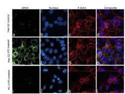
- Immunofluorescence analysis of Beta-2-glycoprotein 1 was performed using 70% confluent log phase Hep G2, Hep G2 treated with PTI (1X for 4hrs) and PC-3 cells treated with PTI (1X for 4hrs). The cells were fixed with 4% paraformaldehyde for 10 minutes, permeabilized with 0.1% Triton™ X-100 for 15 minutes, and blocked with 2% BSA for 45 minutes at room temperature. The cells were labeled with Apolipoprotein H Monoclonal Antibody (4A2F8H12) (Product # MA5-29035) at 1:200 dilution in 0.1% BSA, incubated at 4 degree celsius overnight and then labeled with Donkey anti-Mouse IgG (H+L) Highly Cross-Adsorbed Secondary Antibody, Alexa Fluor Plus 488 (Product # A32766), (1:2000 dilution), for 45 minutes at room temperature (Panel a,e,i : Green). Nuclei (Panel b,f,j :Blue) were stained with ProLong™ Diamond Antifade Mountant with DAPI (Product # P36962). F-actin (Panel c,g,k : Red) was stained with Rhodamine Phalloidin (Product # R415, 1:300 dilution). Panel h represents the merged image showing cytoplasmic localization upon PTI treatment. Panel d and i represents merged images for control cells. The images were captured at 60X magnification.

- Submitted by
- Invitrogen Antibodies (provider)
- Main image

- Experimental details
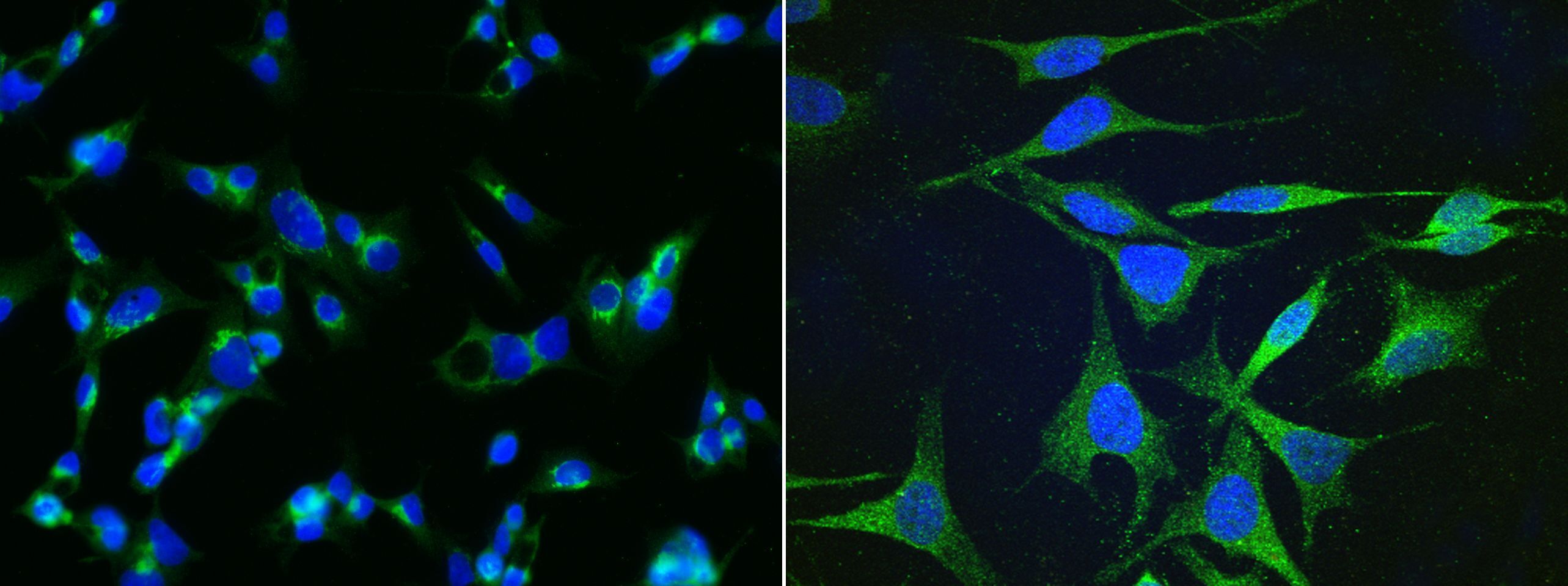
- Immunofluorescence staining of Human Apolipoprotein H in HepG2 or Hela cells. Cells were fixed with 4% PFA, permeabilzed with 1% Triton X-100 in PBS, blocked with 10% serum, and incubated with Apolipoprotein H Monoclonal Antibody (4A2F8H12) (Product # MA5-29035, 1:60). Then cells were stained with the Alexa Fluor® 488-conjugated Goat Anti-mouse IgG secondary antibody (left panel, captured by laser confocal scanning microscope; right panel, captured by fluorescence microscope), counterstained with DAPI (blue). Positive staining was localized to cytoplasm.
- Submitted by
- Invitrogen Antibodies (provider)
- Main image
- Experimental details
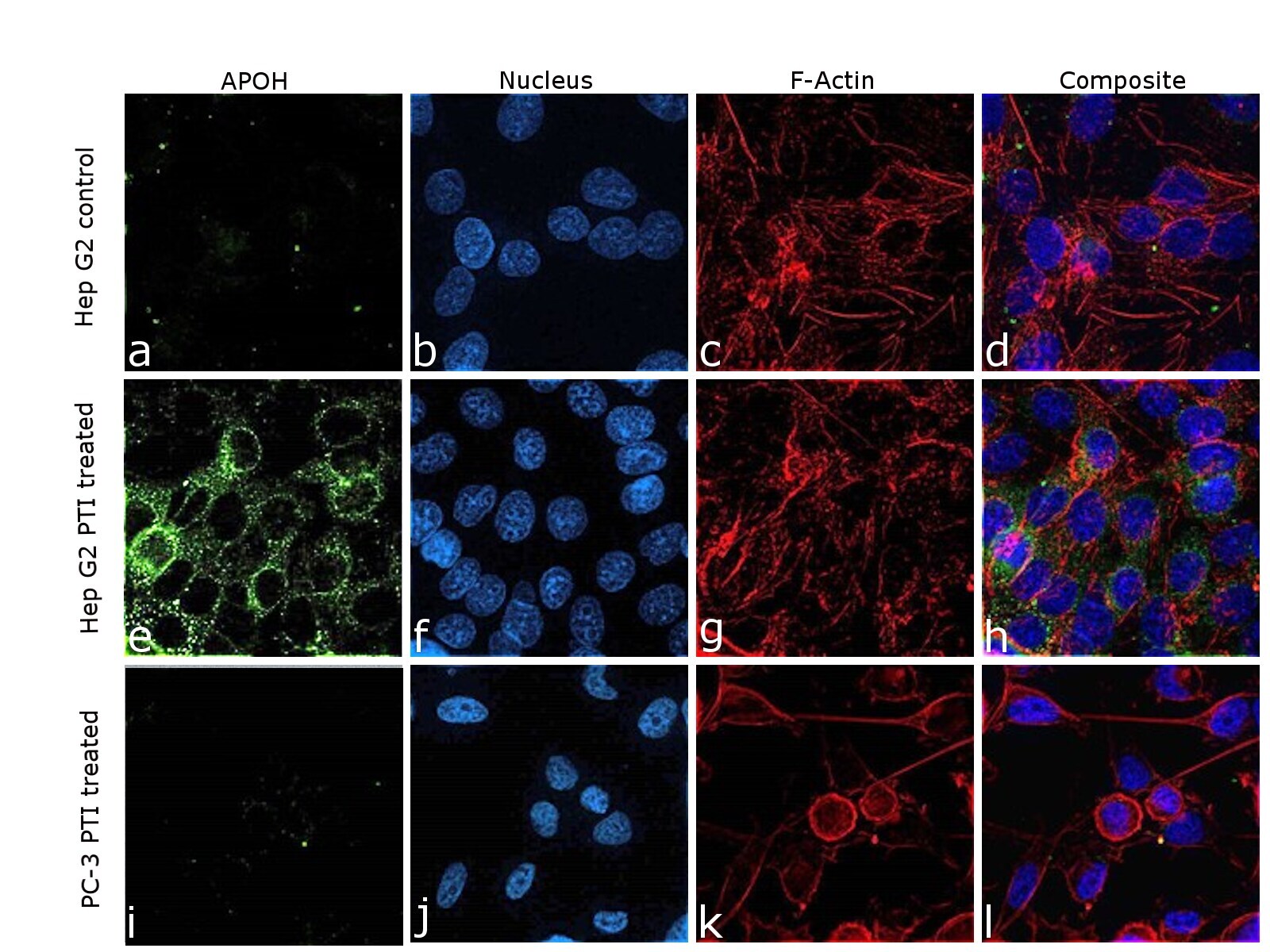
- Immunofluorescence analysis of Beta-2-glycoprotein 1 was performed using 70% confluent log phase Hep G2, Hep G2 treated with PTI (1X for 4hrs) and PC-3 cells treated with PTI (1X for 4hrs). The cells were fixed with 4% paraformaldehyde for 10 minutes, permeabilized with 0.1% Triton™ X-100 for 15 minutes, and blocked with 2% BSA for 45 minutes at room temperature. The cells were labeled with Apolipoprotein H Monoclonal Antibody (4A2F8H12) (Product # MA5-29035) at 1:200 dilution in 0.1% BSA, incubated at 4 degree celsius overnight and then labeled with Donkey anti-Mouse IgG (H+L) Highly Cross-Adsorbed Secondary Antibody, Alexa Fluor Plus 488 (Product # A32766), (1:2000 dilution), for 45 minutes at room temperature (Panel a,e,i : Green). Nuclei (Panel b,f,j :Blue) were stained with ProLong™ Diamond Antifade Mountant with DAPI (Product # P36962). F-actin (Panel c,g,k : Red) was stained with Rhodamine Phalloidin (Product # R415, 1:300 dilution). Panel h represents the merged image showing cytoplasmic localization upon PTI treatment. Panel d and i represents merged images for control cells. The images were captured at 60X magnification.
Supportive validation
- Submitted by
- Invitrogen Antibodies (provider)
- Main image

- Experimental details
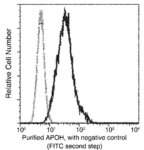
- Flow cytometric analysis of Human Apolipoprotein H expression on MDA-MB-231 cells. The cells were treated according to manufacturer’s manual, stained with Apolipoprotein H Monoclonal Antibody (4A2F8H12) (Product # MA5-29035), then a FITC-conjugated Secondary antibody. The fluorescence histograms were derived from gated events with the forward and side light-scatter characteristics of intact cells.
- Submitted by
- Invitrogen Antibodies (provider)
- Main image

- Experimental details
- Flow cytometric analysis of Human Apolipoprotein H expression on MDA-MB-231 cells. The cells were treated according to manufacturer’s manual, stained with Apolipoprotein H Monoclonal Antibody (4A2F8H12) (Product # MA5-29035), then a FITC-conjugated Secondary antibody. The fluorescence histograms were derived from gated events with the forward and side light-scatter characteristics of intact cells.
