Explore
 Validate
 Learn
Learn Western blot
 Immunocytochemistry
 Immunohistochemistry
Antibody data
- Antibody Data
- Antigen structure
- References [4]
- Comments [0]
- Validations
- Western blot [1]
- Immunocytochemistry [1]
Submit
Validation data
Reference
Comment
Report error
- Product number
- HPA001654 - Provider product page

- Provider
- Atlas Antibodies
- Proper citation
- Atlas Antibodies Cat#HPA001654, RRID:AB_1078181
- Product name
- Anti-APOH
- Antibody type
- Polyclonal
- Description
- Polyclonal Antibody against Human APOH, Gene description: apolipoprotein H (beta-2-glycoprotein I), Alternative Gene Names: B2G1, BG, Validated applications: WB, IHC, ICC, Uniprot ID: P02749, Storage: Store at +4°C for short term storage. Long time storage is recommended at -20°C.
- Reactivity
- Human
- Host
- Rabbit
- Conjugate
- Unconjugated
- Isotype
- IgG
- Vial size
- 100 µl
- Concentration
- 0.1 mg/ml
- Storage
- Store at +4°C for short term storage. Long time storage is recommended at -20°C.
- Handling
- The antibody solution should be gently mixed before use.
Submitted references OxLDL/β2GPI/anti‑β2GPI Ab complex induces inflammatory activation via the TLR4/NF‑κB pathway in HUVECs
β2-Glycoprotein I/HLA class II complexes are novel autoantigens in antiphospholipid syndrome.
Classification of protein profiles from antibody microarrays using heat and detergent treatment
Variance decomposition of protein profiles from antibody arrays using a longitudinal twin model
Zhang G, Cai Q, Zhou H, He C, Chen Y, Zhang P, Wang T, Xu L, Yan J
Molecular Medicine Reports 2020;23(2)
Molecular Medicine Reports 2020;23(2)
β2-Glycoprotein I/HLA class II complexes are novel autoantigens in antiphospholipid syndrome.
Tanimura K, Jin H, Suenaga T, Morikami S, Arase N, Kishida K, Hirayasu K, Kohyama M, Ebina Y, Yasuda S, Horita T, Takasugi K, Ohmura K, Yamamoto K, Katayama I, Sasazuki T, Lanier LL, Atsumi T, Yamada H, Arase H
Blood 2015 Apr 30;125(18):2835-44
Blood 2015 Apr 30;125(18):2835-44
Classification of protein profiles from antibody microarrays using heat and detergent treatment
Häggmark A, Neiman M, Drobin K, Zwahlen M, Uhlén M, Nilsson P, Schwenk J
New Biotechnology 2012;29(5):564-570
New Biotechnology 2012;29(5):564-570
Variance decomposition of protein profiles from antibody arrays using a longitudinal twin model
Kato B, Nicholson G, Neiman M, Rantalainen M, Holmes C, Barrett A, Uhlén M, Nilsson P, Spector T, Schwenk J
Proteome Science 2011;9(1):73
Proteome Science 2011;9(1):73
No comments: Submit comment
Enhanced validation
- Submitted by
- Atlas Antibodies (provider)
- Enhanced method
- Genetic validation
- Main image
- Experimental details
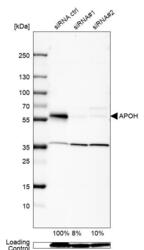
- Western blot analysis in Caco-2 cells transfected with control siRNA, target specific siRNA probe #1 and #2, using Anti-APOH antibody. Remaining relative intensity is presented. Loading control: Anti-GAPDH.
- Sample type
- Human
- Protocol
- Protocol

Supportive validation
- Submitted by
- Atlas Antibodies (provider)
- Main image
- Experimental details
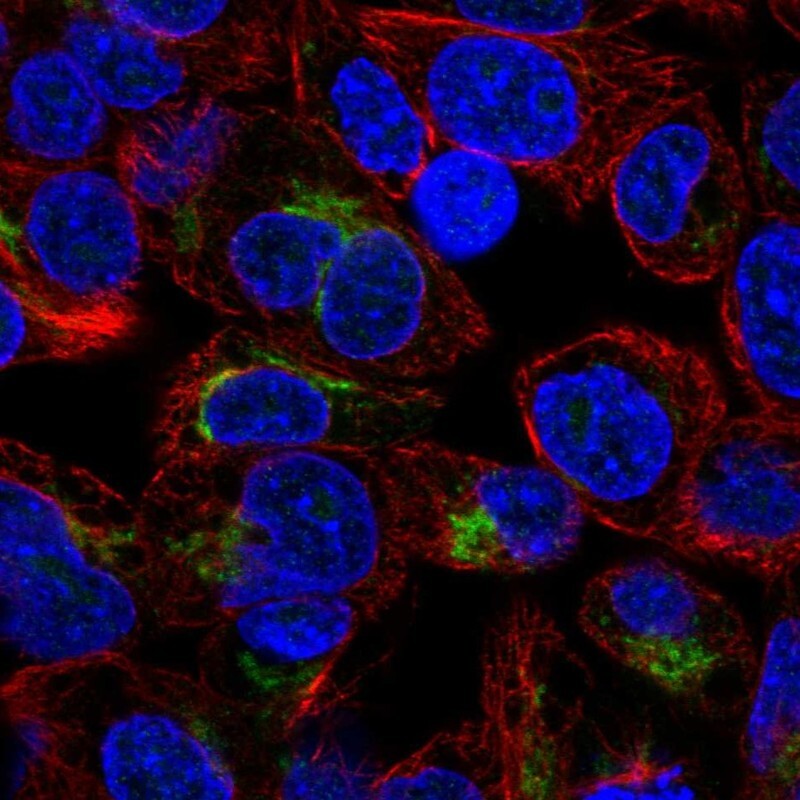
- Immunofluorescent staining of human cell line Hep G2 shows localization to the Golgi apparatus.
- Sample type
- Human