Explore
 Validate
 Learn
Learn Western blot
 Immunocytochemistry
Antibody data
- Antibody Data
- Antigen structure
- References [3]
- Comments [0]
- Validations
- Western blot [2]
- Immunocytochemistry [1]
- Immunohistochemistry [10]
Submit
Validation data
Reference
Comment
Report error
- Product number
- HPA001520 - Provider product page

- Provider
- Atlas Antibodies
- Proper citation
- Atlas Antibodies Cat#HPA001520, RRID:AB_1078243
- Product name
- Anti-ATP5B
- Antibody type
- Polyclonal
- Reactivity
- Human, Mouse, Rat
- Host
- Rabbit
- Conjugate
- Unconjugated
- Antigen sequence
TSPSPKAGAATGRIVAVIGAVVDVQFDEGLPPILN
ALEVQGRETRLVLEVAQHLGESTVRTIAMDGTEGL
VRGQKVLDSGAPIKIPVGPETLGRIMNVIGEPIDE
RGPIKTKQFAPIHAEAPEFMEMSVEQEILVTGIKV
VD- Isotype
- IgG
- Vial size
- 100 µl
- Storage
- Store at +4°C for short term storage. Long time storage is recommended at -20°C.
Submitted references C11orf83, a mitochondrial cardiolipin-binding protein involved in bc1 complex assembly and supercomplex stabilization.
Surrogate antigens as targets for proteome-wide binder selection
Resveratrol induces SIRT1- and energy–stress-independent inhibition of tumor cell regrowth after low-dose platinum treatment
Desmurs M, Foti M, Raemy E, Vaz FM, Martinou JC, Bairoch A, Lane L
Molecular and cellular biology 2015 Apr;35(7):1139-56
Molecular and cellular biology 2015 Apr;35(7):1139-56
Surrogate antigens as targets for proteome-wide binder selection
Gustavsson E, Ek S, Steen J, Kristensson M, Älgenäs C, Uhlén M, Wingren C, Ottosson J, Hober S, Borrebaeck C
New Biotechnology 2011 July;28(4):302-311
New Biotechnology 2011 July;28(4):302-311
Resveratrol induces SIRT1- and energy–stress-independent inhibition of tumor cell regrowth after low-dose platinum treatment
Björklund M, Roos J, Gogvadze V, Shoshan M
Cancer Chemotherapy and Pharmacology 2011 December;68(6):1459-1467
Cancer Chemotherapy and Pharmacology 2011 December;68(6):1459-1467
No comments: Submit comment
Enhanced validation
- Submitted by
- Atlas Antibodies (provider)
- Enhanced method
- Genetic validation
- Main image

- Experimental details
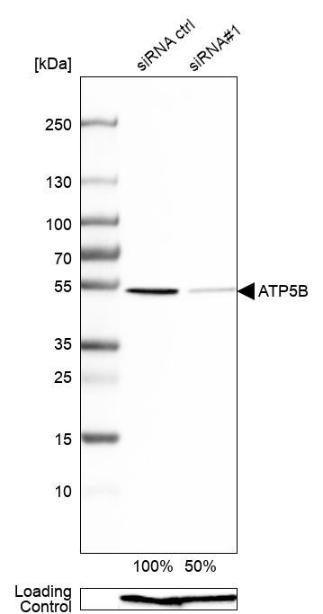
- Western blot analysis in A-549 cells transfected with control siRNA, target specific siRNA probe #1, using Anti-ATP5B antibody. Remaining relative intensity is presented. Loading control: Anti-GAPDH.
- Submitted by
- Atlas Antibodies (provider)
- Enhanced method
- Independent antibody validation
- Main image

- Experimental details
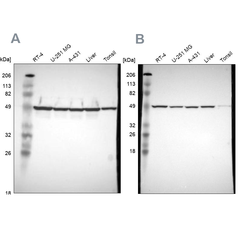
- Western blot analysis using Anti-ATP5B antibody HPA001520 (A) shows similar pattern to independent antibody HPA001528 (B).
Supportive validation
- Submitted by
- Atlas Antibodies (provider)
- Main image

- Experimental details
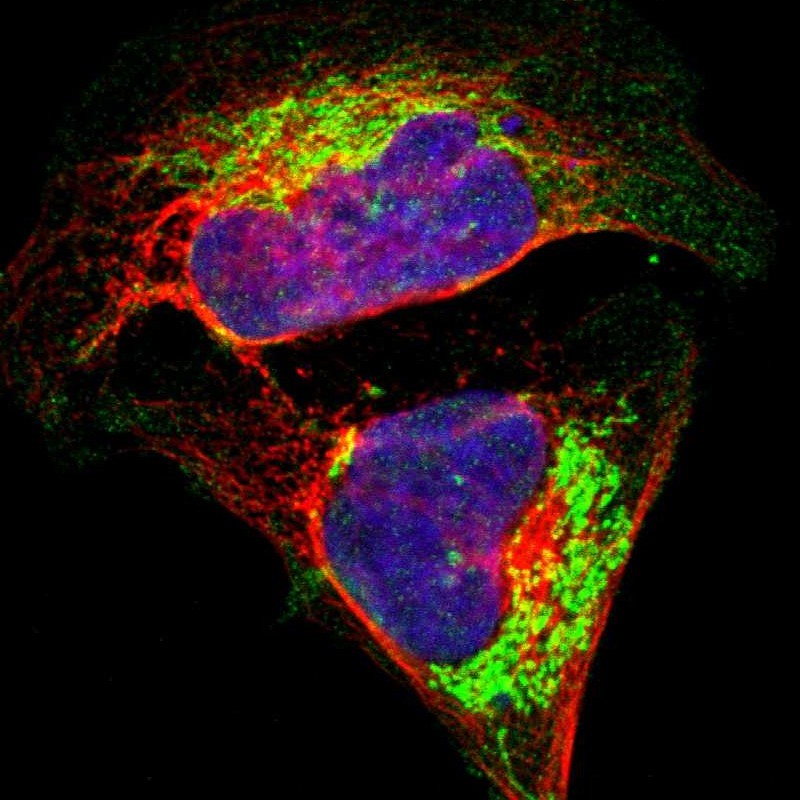
- Immunofluorescent staining of human cell line U-2 OS shows localization to mitochondria.
- Sample type
- HUMAN
Enhanced validation
Supportive validation
- Submitted by
- Atlas Antibodies (provider)
- Enhanced method
- Independent antibody validation
- Main image
- Experimental details
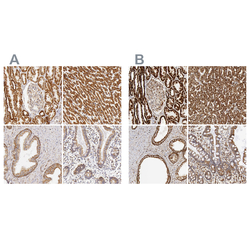
- Immunohistochemical staining of human kidney, liver, prostate and small intestine using Anti-ATP5B antibody HPA001520 (A) shows similar protein distribution across tissues to independent antibody HPA001528 (B).

Supportive validation
- Submitted by
- Atlas Antibodies (provider)
- Main image
- Experimental details
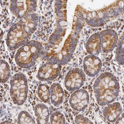
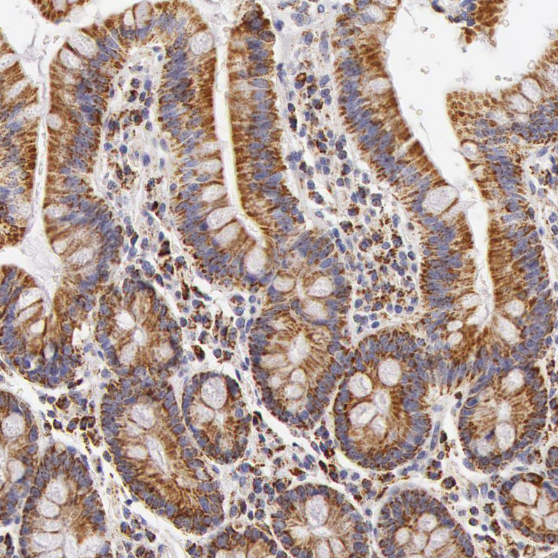
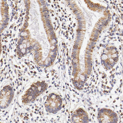
- Immunohistochemical staining of human duodenum shows strong cytoplasmic positivity in glandular cells.

- Submitted by
- Atlas Antibodies (provider)
- Main image
- Experimental details
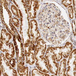
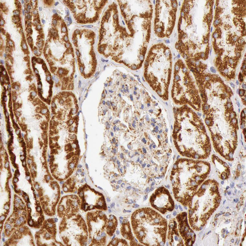
- Immunohistochemical staining of human kidney shows strong granular cytoplasmic positivity in cells in tubules and moderate immunoreactivity in glomeruli.
- Sample type
- HUMAN

- Submitted by
- Atlas Antibodies (provider)
- Main image
- Experimental details
- Immunohistochemical staining of human duodenum shows moderate granular cytoplasmic positivity in glandular cells.
- Sample type
- HUMAN
- Submitted by
- Atlas Antibodies (provider)
- Main image
- Experimental details
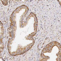
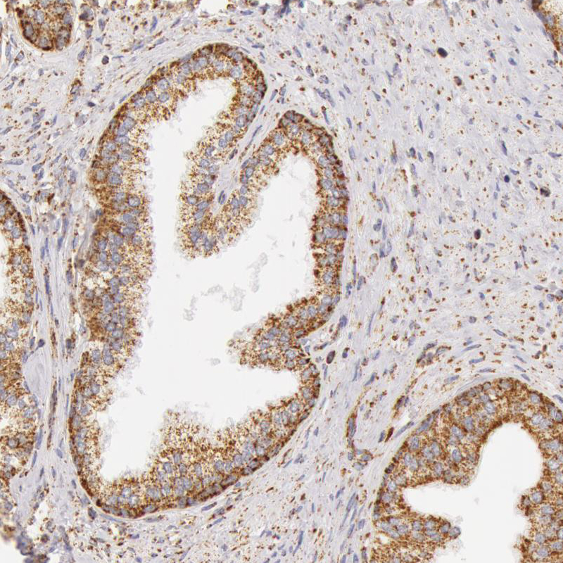
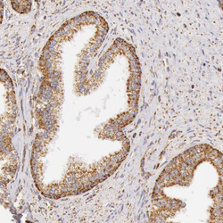
- Immunohistochemical staining of human prostate shows moderate granular cytoplasmic positivity in glandular cells.
- Sample type
- HUMAN
- Submitted by
- Atlas Antibodies (provider)
- Main image
- Experimental details
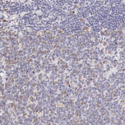
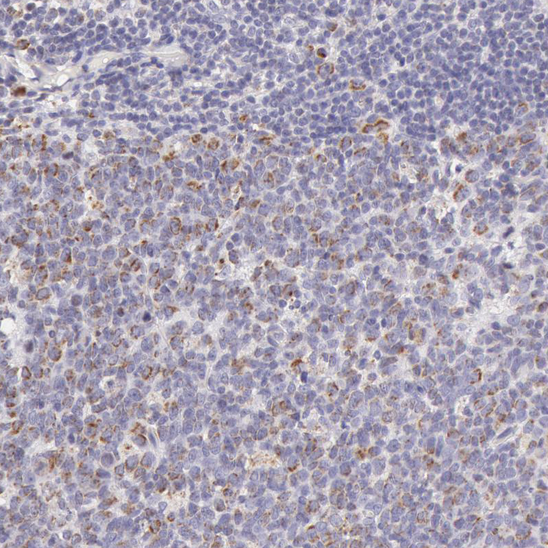
- Immunohistochemical staining of human tonsil shows weak granular cytoplasmic positivity in lymphoid cells, mainly in reaction centrum.
- Sample type
- HUMAN
- Submitted by
- Atlas Antibodies (provider)
- Main image
- Experimental details
- Immunohistochemical staining of human prostate shows moderate granular cytoplasmic positivity in glandular cells.
- Sample type
- HUMAN

- Submitted by
- Atlas Antibodies (provider)
- Main image
- Experimental details
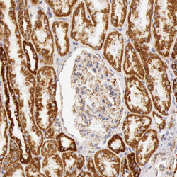
- Immunohistochemical staining of human kidney shows moderate granular cytoplasmic positivity in cells in tubules.
- Sample type
- HUMAN
- Submitted by
- Atlas Antibodies (provider)
- Main image
- Experimental details
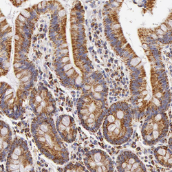
- Immunohistochemical staining of human small intestine shows moderate granular cytoplasmic positivity in glandular cells.
- Sample type
- HUMAN

- Submitted by
- Atlas Antibodies (provider)
- Main image
- Experimental details
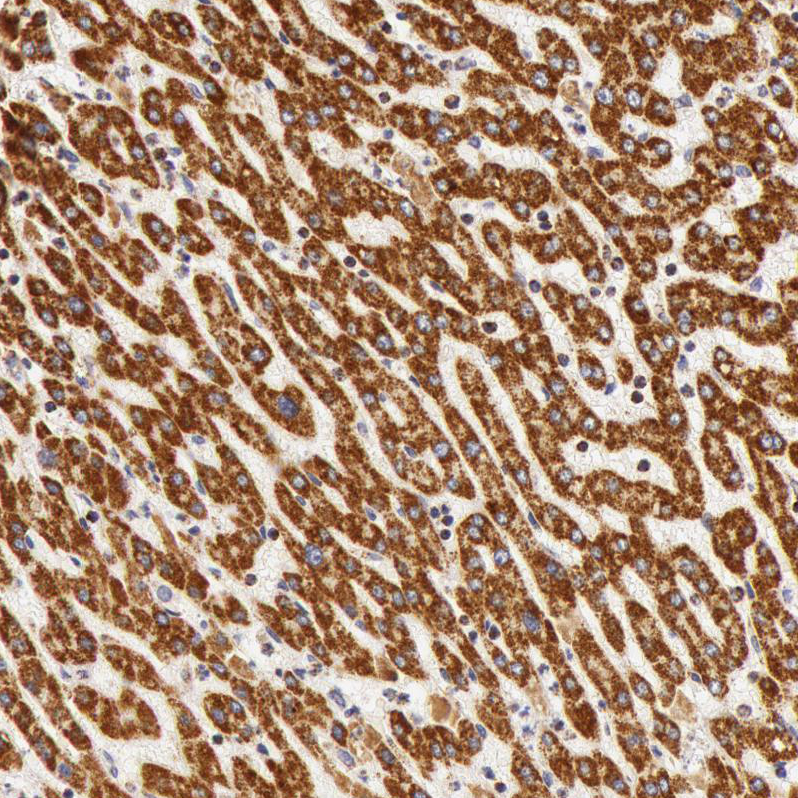
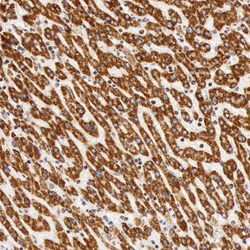
- Immunohistochemical staining of human liver shows strong granular cytoplasmic positivity in hepatocytes.
- Sample type
- HUMAN