Explore
 Validate
 Learn
Learn Western blot
 Immunocytochemistry
Antibody data
- Antibody Data
- Antigen structure
- References [11]
- Comments [0]
- Validations
- Immunocytochemistry [10]
- Immunoprecipitation [1]
- Other assay [4]
Submit
Validation data
Reference
Comment
Report error
- Product number
- MA1-930 - Provider product page

- Provider
- Invitrogen Antibodies
- Product name
- ATP Synthase beta Monoclonal Antibody (4.3E8.D10)
- Antibody type
- Monoclonal
- Antigen
- Other
- Description
- MA1-930 detects the beta subunit of ATP synthase from mouse rat and human samples. This antibody is useful as a mitochondrial marker. MA1-930 has been successfully used in immunofluorescence, western blot, and immunoprecipitation procedures. By immunoprecipitation, this antibody detects an 50 kDa protein representing ATP synthase from solubilized rat brain mitochondria.
- Reactivity
- Human, Mouse, Rat
- Host
- Mouse
- Isotype
- IgG
- Antibody clone number
- 4.3E8.D10
- Vial size
- 100 μg
- Concentration
- 1 mg/mL
- Storage
- -20°C, Avoid Freeze/Thaw Cycles
Submitted references Metabolic Adaptation of Macrophages as Mechanism of Defense against Crystalline Silica.
Enhancing Proteotoxic Stress in Leiomyosarcoma Cells Triggers Mitochondrial Dysfunctions, Cell Death, and Antitumor Activity in vivo.
CARD19, the protein formerly known as BinCARD, is a mitochondrial protein that does not regulate Bcl10-dependent NF-κB activation after TCR engagement.
ACE overexpression in myeloid cells increases oxidative metabolism and cellular ATP.
Age-dependent changes in intervertebral disc cell mitochondria and bioenergetics.
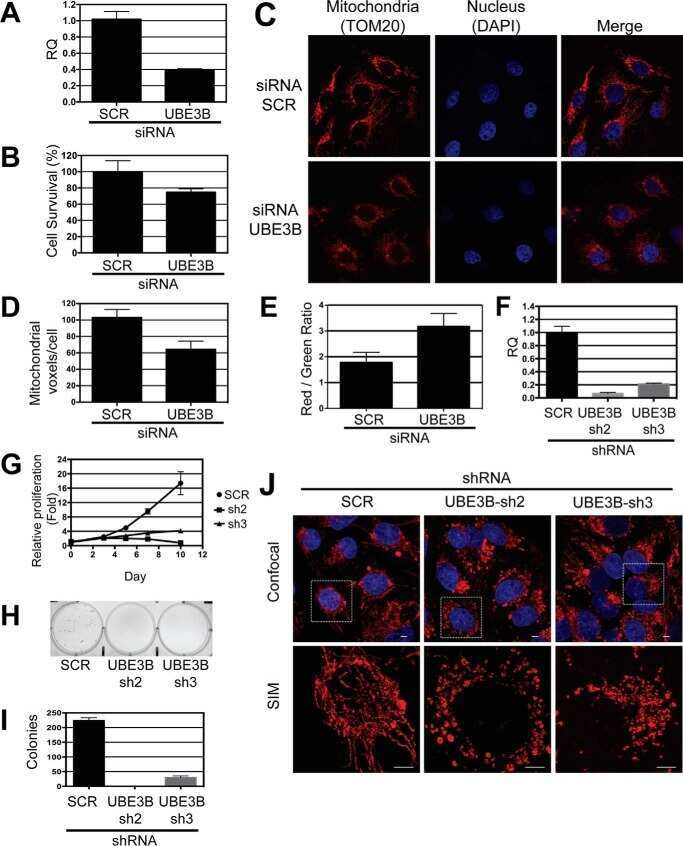
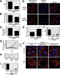
UBE3B Is a Calmodulin-regulated, Mitochondrion-associated E3 Ubiquitin Ligase.
PINK1 deficiency impairs mitochondrial homeostasis and promotes lung fibrosis.
Human brain endothelial ATP synthase beta-subunit is mannose-insensitive binding target of FimH.
Expression and subcellular localization of BNIP3 in hypoxic hepatocytes and liver stress.
The Krebs cycle and mitochondrial mass are early victims of endothelial dysfunction: proteomic approach.
Polyamine transport by mammalian cells and mitochondria: role of antizyme and glycosaminoglycans.
Marrocco A, Frawley K, Pearce LL, Peterson J, O'Brien JP, Mullett SJ, Wendell SG, St Croix CM, Mischler SE, Ortiz LA
Journal of immunology (Baltimore, Md. : 1950) 2021 Sep 15;207(6):1627-1640
Journal of immunology (Baltimore, Md. : 1950) 2021 Sep 15;207(6):1627-1640
Enhancing Proteotoxic Stress in Leiomyosarcoma Cells Triggers Mitochondrial Dysfunctions, Cell Death, and Antitumor Activity in vivo.
Iuliano L, Drioli S, Pignochino Y, Cafiero CM, Minisini M, D'Este F, Picco R, Dalla E, Giordano G, Grignani G, Di Giorgio E, Benedetti F, Felluga F, Brancolini C
Molecular cancer therapeutics 2021 Jun;20(6):1039-1051
Molecular cancer therapeutics 2021 Jun;20(6):1039-1051
CARD19, the protein formerly known as BinCARD, is a mitochondrial protein that does not regulate Bcl10-dependent NF-κB activation after TCR engagement.
Rios KE, Kashyap AK, Maynard SK, Washington M, Paul S, Schaefer BC
Cellular immunology 2020 Oct;356:104179
Cellular immunology 2020 Oct;356:104179
ACE overexpression in myeloid cells increases oxidative metabolism and cellular ATP.
Cao DY, Spivia WR, Veiras LC, Khan Z, Peng Z, Jones AE, Bernstein EA, Saito S, Okwan-Duodu D, Parker SJ, Giani JF, Divakaruni AS, Van Eyk JE, Bernstein KE
The Journal of biological chemistry 2020 Jan 31;295(5):1369-1384
The Journal of biological chemistry 2020 Jan 31;295(5):1369-1384
Age-dependent changes in intervertebral disc cell mitochondria and bioenergetics.
Hartman R, Patil P, Tisherman R, St Croix C, Niedernhofer LJ, Robbins PD, Ambrosio F, Van Houten B, Sowa G, Vo N
European cells & materials 2018 Oct 18;36:171-183
European cells & materials 2018 Oct 18;36:171-183
UBE3B Is a Calmodulin-regulated, Mitochondrion-associated E3 Ubiquitin Ligase.
Braganza A, Li J, Zeng X, Yates NA, Dey NB, Andrews J, Clark J, Zamani L, Wang XH, St Croix C, O'Sullivan R, Garcia-Exposito L, Brodsky JL, Sobol RW
The Journal of biological chemistry 2017 Feb 10;292(6):2470-2484
The Journal of biological chemistry 2017 Feb 10;292(6):2470-2484
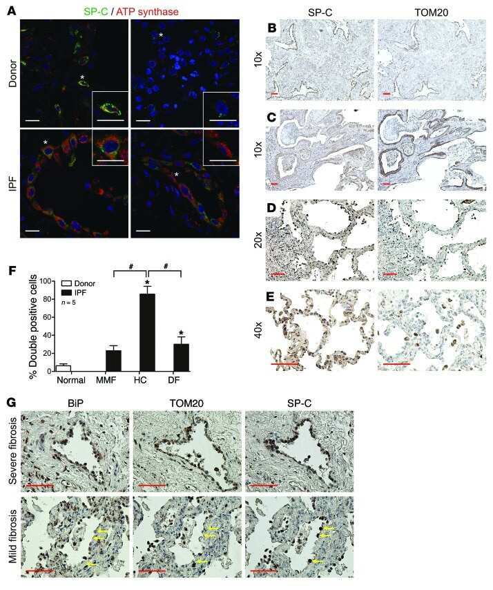
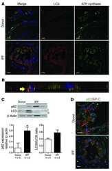
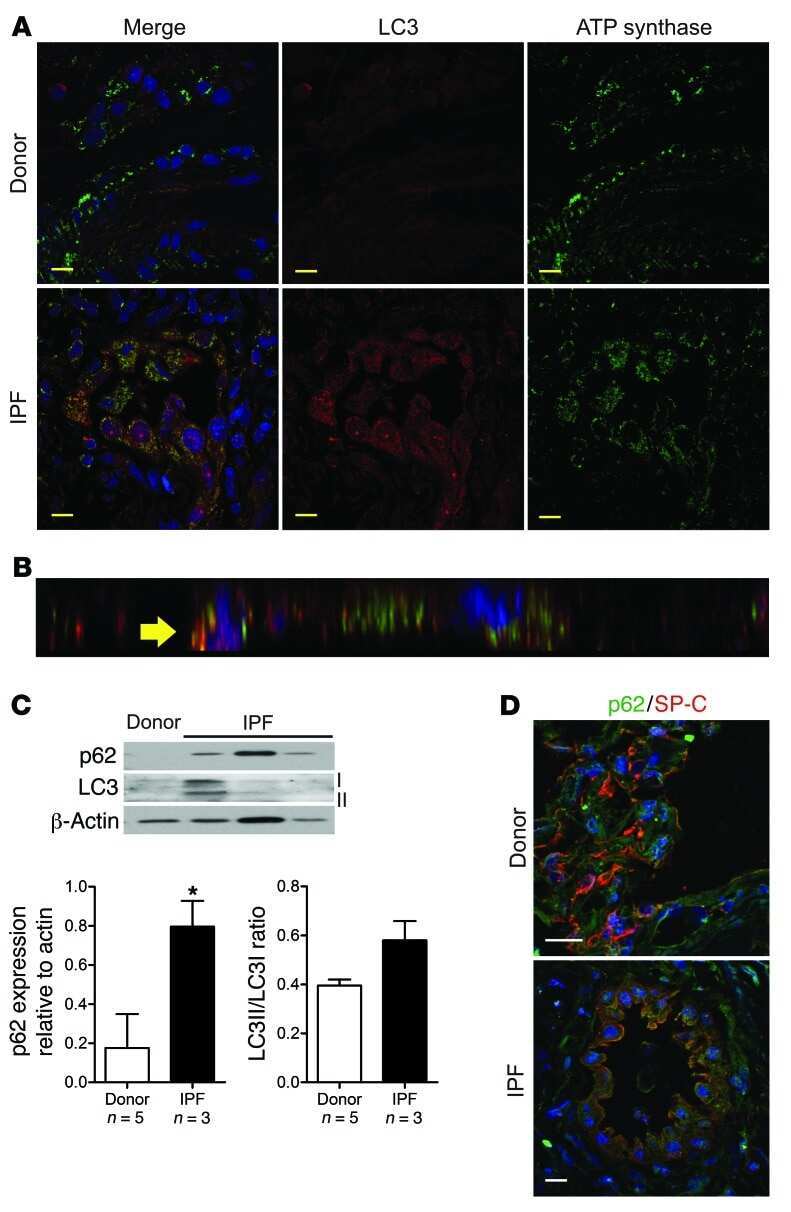
PINK1 deficiency impairs mitochondrial homeostasis and promotes lung fibrosis.
Bueno M, Lai YC, Romero Y, Brands J, St Croix CM, Kamga C, Corey C, Herazo-Maya JD, Sembrat J, Lee JS, Duncan SR, Rojas M, Shiva S, Chu CT, Mora AL
The Journal of clinical investigation 2015 Feb;125(2):521-38
The Journal of clinical investigation 2015 Feb;125(2):521-38
Human brain endothelial ATP synthase beta-subunit is mannose-insensitive binding target of FimH.
Shin S, Kim KS
FEMS microbiology letters 2010 Feb;303(2):156-62
FEMS microbiology letters 2010 Feb;303(2):156-62
Expression and subcellular localization of BNIP3 in hypoxic hepatocytes and liver stress.
Metukuri MR, Beer-Stolz D, Namas RA, Dhupar R, Torres A, Loughran PA, Jefferson BS, Tsung A, Billiar TR, Vodovotz Y, Zamora R
American journal of physiology. Gastrointestinal and liver physiology 2009 Mar;296(3):G499-509
American journal of physiology. Gastrointestinal and liver physiology 2009 Mar;296(3):G499-509
The Krebs cycle and mitochondrial mass are early victims of endothelial dysfunction: proteomic approach.
Addabbo F, Ratliff B, Park HC, Kuo MC, Ungvari Z, Csiszar A, Krasnikov B, Sodhi K, Zhang F, Nasjletti A, Goligorsky MS
The American journal of pathology 2009 Jan;174(1):34-43
The American journal of pathology 2009 Jan;174(1):34-43
Polyamine transport by mammalian cells and mitochondria: role of antizyme and glycosaminoglycans.
Hoshino K, Momiyama E, Yoshida K, Nishimura K, Sakai S, Toida T, Kashiwagi K, Igarashi K
The Journal of biological chemistry 2005 Dec 30;280(52):42801-8
The Journal of biological chemistry 2005 Dec 30;280(52):42801-8
No comments: Submit comment
Supportive validation
- Submitted by
- Invitrogen Antibodies (provider)
- Main image

- Experimental details
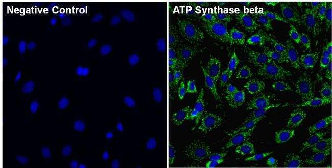
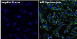
- Immunofluorescent analysis of ATP Synthase beta (green) in 3T3 cells. The cells were fixed with 4% paraformaldehyde for 15 minutes and blocked with 3% Blocker BSA (Product # 37525) in PBS for 15 minutes at room temperature. Cells were stained with or without ATP Synthase beta mouse monoclonal antibody (Product # MA1-930), at a concentration of 5 µg/mL for 1 hour at room temperature, and then incubated with a Goat anti-Mouse IgG (H+L) Superclonal Secondary Antibody, Alexa Fluor® 488 conjugate (Product # A28175) at a dilution of 1:1000 for 1 hour at room temperature (both panels, green). Nuclei (both panels, blue) were stained with Hoechst 33342 dye (Product # 62249). Images were taken on a Thermo Scientific ToxInsight at 20X magnification.
- Submitted by
- Invitrogen Antibodies (provider)
- Main image
- Experimental details
- Immunofluorescent analysis of ATP Synthase beta (red) in HEK293T cells. Cells fixed with 4% formaldehyde were permeabilized and blocked with 1X PBS containing 5% BSA and 0.3% Triton X-100 for 1 hour at room temperature. Cells were probed with an ATP Synthase beta monoclonal antibody (Product # MA1-930) at a dilution of 1:100 overnight at 4°C in 1X PBS containing 1% BSA and 0.3% Triton X-100, washed with 1X PBS, and incubated with a fluorophore-conjugated goat anti-mouse IgG secondary antibody at a dilution of 1:200 for 1 hour at room temperature. Nuclei (blue) were stained with DAPI. Images were taken on a Leica DM1000 microscope at 40X magnification. Data courtesy of the Innovators Program.

- Submitted by
- Invitrogen Antibodies (provider)
- Main image

- Experimental details
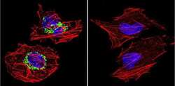
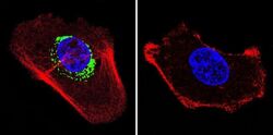
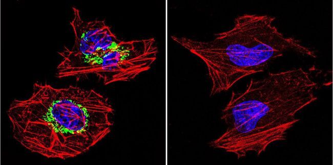
- Immunofluorescent analysis of ATP Synthase beta in A431 cells. Cells were grown on chamber slides and fixed with formaldehyde prior to staining. Cells were probed without (control) or with a ATP Synthase beta monoclonal antibody (Product # MA1-930) at a dilution of 1:200 overnight at 4 C, washed with PBS and incubated with a DyLight-488 conjugated secondary antibody (Product # 35503). ATP Synthase beta staining (green), F-Actin staining with Phalloidin (red) and nuclei with DAPI (blue) is shown. Images were taken at 60X magnification.

- Submitted by
- Invitrogen Antibodies (provider)
- Main image
- Experimental details
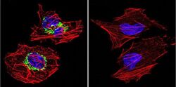
- Immunofluorescent analysis of ATP Synthase beta in HeLa cells. Cells were grown on chamber slides and fixed with formaldehyde prior to staining. Cells were probed without (control) or with a ATP Synthase beta monoclonal antibody (Product # MA1-930) at a dilution of 1:200 overnight at 4 C, washed with PBS and incubated with a DyLight-488 conjugated secondary antibody (Product # 35503). ATP Synthase beta staining (green), F-Actin staining with Phalloidin (red) and nuclei with DAPI (blue) is shown. Images were taken at 60X magnification.

- Submitted by
- Invitrogen Antibodies (provider)
- Main image
- Experimental details
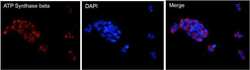
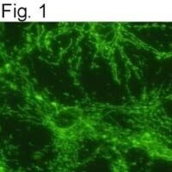
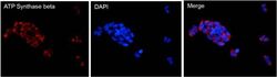
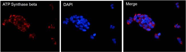
- Indirect immunofluorescence staining of rat neuronal/glial cell culture using Product # MA1-930.
- Submitted by
- Invitrogen Antibodies (provider)
- Main image
- Experimental details
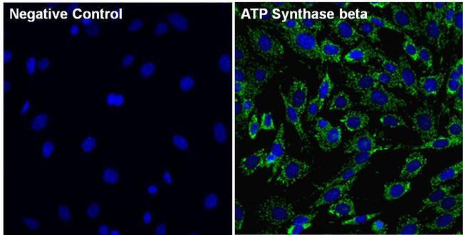
- Immunofluorescent analysis of ATP Synthase beta (green) in 3T3 cells. The cells were fixed with 4% paraformaldehyde for 15 minutes and blocked with 3% Blocker BSA (Product # 37525) in PBS for 15 minutes at room temperature. Cells were stained with or without ATP Synthase beta mouse monoclonal antibody (Product # MA1-930), at a concentration of 5 µg/mL for 1 hour at room temperature, and then incubated with a Goat anti-Mouse IgG (H+L) Superclonal Secondary Antibody, Alexa Fluor® 488 conjugate (Product # A28175) at a dilution of 1:1000 for 1 hour at room temperature (both panels, green). Nuclei (both panels, blue) were stained with Hoechst 33342 dye (Product # 62249). Images were taken on a Thermo Scientific ToxInsight at 20X magnification.
- Submitted by
- Invitrogen Antibodies (provider)
- Main image

- Experimental details
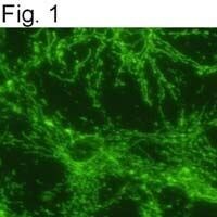
- Indirect immunofluorescence staining of rat neuronal/glial cell culture using Product # MA1-930.

- Submitted by
- Invitrogen Antibodies (provider)
- Main image
- Experimental details
- Immunofluorescent analysis of ATP Synthase beta (red) in HEK293T cells. Cells fixed with 4% formaldehyde were permeabilized and blocked with 1X PBS containing 5% BSA and 0.3% Triton X-100 for 1 hour at room temperature. Cells were probed with an ATP Synthase beta monoclonal antibody (Product # MA1-930) at a dilution of 1:100 overnight at 4°C in 1X PBS containing 1% BSA and 0.3% Triton X-100, washed with 1X PBS, and incubated with a fluorophore-conjugated goat anti-mouse IgG secondary antibody at a dilution of 1:200 for 1 hour at room temperature. Nuclei (blue) were stained with DAPI. Images were taken on a Leica DM1000 microscope at 40X magnification. Data courtesy of the Innovators Program.
- Submitted by
- Invitrogen Antibodies (provider)
- Main image
- Experimental details
- Immunofluorescent analysis of ATP Synthase beta in A431 cells. Cells were grown on chamber slides and fixed with formaldehyde prior to staining. Cells were probed without (control) or with a ATP Synthase beta monoclonal antibody (Product # MA1-930) at a dilution of 1:200 overnight at 4 C, washed with PBS and incubated with a DyLight-488 conjugated secondary antibody (Product # 35503). ATP Synthase beta staining (green), F-Actin staining with Phalloidin (red) and nuclei with DAPI (blue) is shown. Images were taken at 60X magnification.

- Submitted by
- Invitrogen Antibodies (provider)
- Main image
- Experimental details
- Immunofluorescent analysis of ATP Synthase beta in HeLa cells. Cells were grown on chamber slides and fixed with formaldehyde prior to staining. Cells were probed without (control) or with a ATP Synthase beta monoclonal antibody (Product # MA1-930) at a dilution of 1:200 overnight at 4 C, washed with PBS and incubated with a DyLight-488 conjugated secondary antibody (Product # 35503). ATP Synthase beta staining (green), F-Actin staining with Phalloidin (red) and nuclei with DAPI (blue) is shown. Images were taken at 60X magnification.
Supportive validation
- Submitted by
- Invitrogen Antibodies (provider)
- Main image
- Experimental details
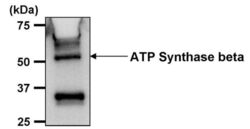
- Immunoprecipitation of ATP Synthase beta was performed on THP-1 cells. Antigen-antibody complexes were formed by incubating 500 µg of THP-1 whole cell lysate (in 500 µL volume) with 5 µL of an ATP Synthase beta monoclonal antibody (Product # MA1-930) overnight at 4°C. The immune complexes were captured on 30 µL of protein G agarose, washed extensively, and eluted with 6X Laemmli non-reducing sample buffer. Samples were resolved on an 8% SDS-PAGE gel, transferred to a PVDF membrane, and blocked with 5% milk in TBST for 1 hour at room temperature. The membrane was probed with an ATP Synthase beta monoclonal antibody (Product # MA1-930) at a dilution of 1:1000 overnight at 4°C, washed in TBST, and probed with an HRP-conjugated goat anti-mouse IgG secondary at a dilution of 1:40,000 for 1 hour at room temperature. Chemiluminescent detection was performed using ECL substrate. Data courtesy of the Innovators Program.

Supportive validation
- Submitted by
- Invitrogen Antibodies (provider)
- Main image

- Experimental details
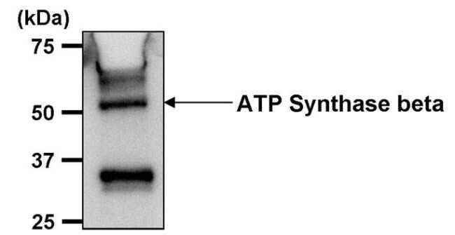
- Immunoprecipitation of ATP Synthase beta was performed on THP-1 cells. Antigen-antibody complexes were formed by incubating 500 µg of THP-1 whole cell lysate (in 500 µL volume) with 5 µL of an ATP Synthase beta monoclonal antibody (Product # MA1-930) overnight at 4°C. The immune complexes were captured on 30 µL of protein G agarose, washed extensively, and eluted with 6X Laemmli non-reducing sample buffer. Samples were resolved on an 8% SDS-PAGE gel, transferred to a PVDF membrane, and blocked with 5% milk in TBST for 1 hour at room temperature. The membrane was probed with an ATP Synthase beta monoclonal antibody (Product # MA1-930) at a dilution of 1:1000 overnight at 4øC, washed in TBST, and probed with an HRP-conjugated goat anti-mouse IgG secondary at a dilution of 1:40,000 for 1 hour at room temperature. Chemiluminescent detection was performed using ECL substrate. Data courtesy of the Innovators Program.
- Submitted by
- Invitrogen Antibodies (provider)
- Main image
- Experimental details
- NULL
- Submitted by
- Invitrogen Antibodies (provider)
- Main image
- Experimental details
- NULL
- Submitted by
- Invitrogen Antibodies (provider)
- Main image
- Experimental details
- NULL