Explore
 Validate
 Learn
Learn Western blot
 Immunohistochemistry
Antibody data
- Antibody Data
- Antigen structure
- References [2]
- Comments [0]
- Validations
- Immunohistochemistry [1]
Submit
Validation data
Reference
Comment
Report error
- Product number
- PB9869 - Provider product page

- Provider
- Boster Biological Technology
- Product name
- Anti-VEGFD/FIGF Antibody Picoband™
- Antibody type
- Polyclonal
- Description
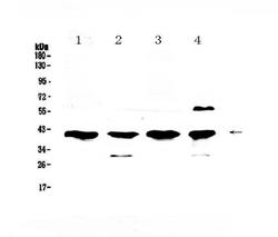
- Polyclonal antibody for FIGF detection. Host: Rabbit.Size: 100μg/vial. Tested applications: IHC-P. Reactive species: Human. FIGF information: Molecular Weight: 40444 MW; Subcellular Localization: Secreted; Tissue Specificity: Highly expressed in lung, heart, small intestine and fetal lung, and at lower levels in skeletal muscle, colon, and pancreas.
- Reactivity
- Human, Mouse, Rat
- Host
- Rabbit
- Vial size
- 100μg/vial
- Concentration
- Add 0.2ml of distilled water will yield a concentration of 500ug/ml.
- Storage
- At -20°C for one year. After reconstitution, at 4°C for one month. It can also be aliquoted and stored frozen at -20°C for a longer time. Avoid repeated freezing and thawing.
- Handling
- Add 0.2ml of distilled water will yield a concentration of 500ug/ml.
Submitted references VEGF-D-induced draining lymphatic enlargement and tumor lymphangiogenesis promote lymph node metastasis in a xenograft model of ovarian carcinoma.
Efficacy and safety of (32)P-nanocolloid for treatment of distant lymph node metastasis in VX2 tumor-bearing rabbits.
Du LC, Chen XC, Wang D, Wen YJ, Wang CT, Wang XM, Kan B, Wei YQ, Zhao X
Reproductive biology and endocrinology : RB&E 2014 Feb 6;12:14
Reproductive biology and endocrinology : RB&E 2014 Feb 6;12:14
Efficacy and safety of (32)P-nanocolloid for treatment of distant lymph node metastasis in VX2 tumor-bearing rabbits.
Dong S, Huang G, Liu P, Ma Y, Yan W, Wan L, Zhu C
Annals of nuclear medicine 2008 Dec;22(10):849-58
Annals of nuclear medicine 2008 Dec;22(10):849-58
No comments: Submit comment
Supportive validation
- Submitted by
- Boster Biological Technology (provider)
- Main image
- Experimental details
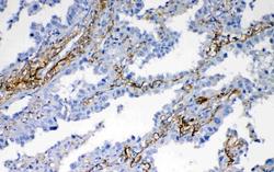
- IHC analysis of VEGFD using anti-VEGFD antibody (PB9869). VEGFD was detected in paraffin-embedded section of human lung cancer tissue. Heat mediated antigen retrieval was performed in citrate buffer (pH6, epitope retrieval solution) for 20 mins. The tissue section was blocked with 10% goat serum. The tissue section was then incubated with 1μg/ml rabbit anti-VEGFD Antibody (PB9869) overnight at 4°C. Biotinylated goat anti-rabbit IgG was used as secondary antibody and incubated for 30 minutes at 37°C. The tissue section was developed using Strepavidin-Biotin-Complex (SABC)(Catalog # SA1022) with DAB as the chromogen.
- Additional image