Explore
 Validate
 Learn
Learn Immunohistochemistry
Antibody data
- Antibody Data
- Antigen structure
- References [4]
- Comments [0]
- Validations
- Immunohistochemistry [1]
Submit
Validation data
Reference
Comment
Report error
- Product number
- HPA001804 - Provider product page

- Provider
- Atlas Antibodies
- Proper citation
- Atlas Antibodies Cat#HPA001804, RRID:AB_1078792
- Product name
- Anti-F13A1
- Antibody type
- Polyclonal
- Description
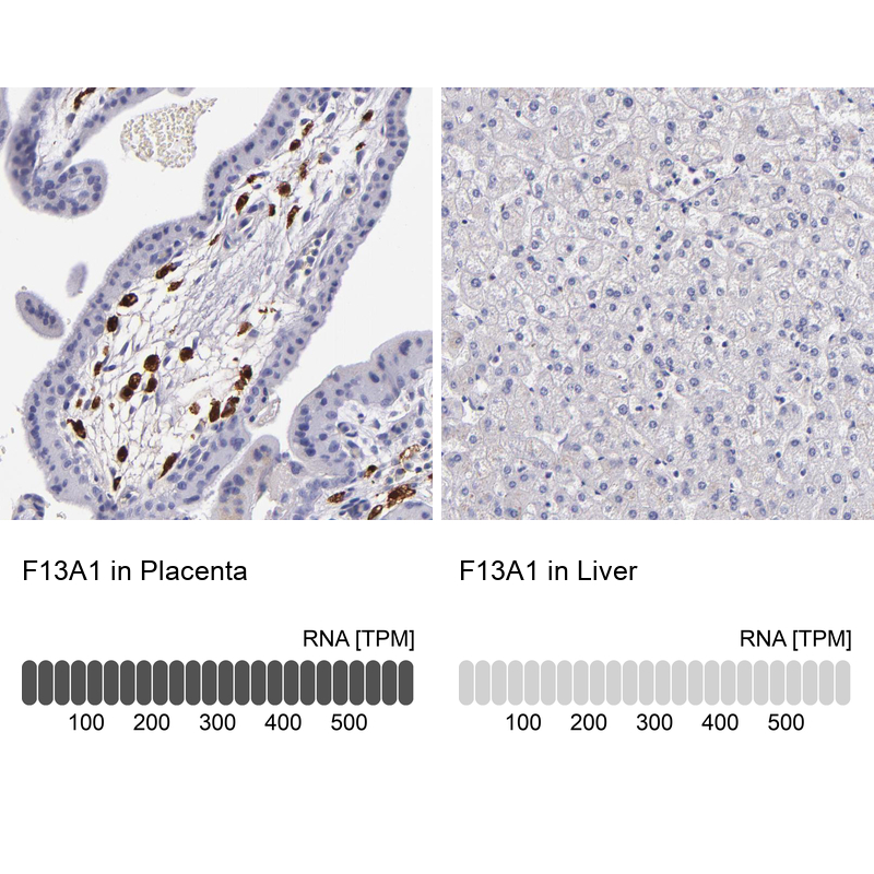
- Polyclonal Antibody against Human F13A1, Gene description: coagulation factor XIII, A1 polypeptide, Alternative Gene Names: F13A, Validated applications: IHC, Uniprot ID: P00488, Storage: Store at +4°C for short term storage. Long time storage is recommended at -20°C.
- Reactivity
- Human
- Host
- Rabbit
- Conjugate
- Unconjugated
- Isotype
- IgG
- Vial size
- 100 µl
- Concentration
- 0.1 mg/ml
- Storage
- Store at +4°C for short term storage. Long time storage is recommended at -20°C.
- Handling
- The antibody solution should be gently mixed before use.
Submitted references Macrophage M2 Co-expression Factors Correlate With the Immune Microenvironment and Predict Outcome of Renal Clear Cell Carcinoma
Genetic determinants of circulating GIP and GLP-1 concentrations
Proteomics analysis of melanoma metastases: association between S100A13 expression and chemotherapy resistance
Variance decomposition of protein profiles from antibody arrays using a longitudinal twin model
Wang Y, Yan K, Lin J, Li J, Bi J
Frontiers in Genetics 2021;12
Frontiers in Genetics 2021;12
Genetic determinants of circulating GIP and GLP-1 concentrations
Almgren P, Lindqvist A, Krus U, Hakaste L, Ottosson-Laakso E, Asplund O, Sonestedt E, Prasad R, Laurila E, Orho-Melander M, Melander O, Tuomi T, Holst J, Nilsson P, Wierup N, Groop L, Ahlqvist E
JCI Insight 2017;2(21)
JCI Insight 2017;2(21)
Proteomics analysis of melanoma metastases: association between S100A13 expression and chemotherapy resistance
Azimi A, Pernemalm M, Frostvik Stolt M, Hansson J, Lehtiö J, Egyházi Brage S, Hertzman Johansson C
British Journal of Cancer 2014;110(10):2489-2495
British Journal of Cancer 2014;110(10):2489-2495
Variance decomposition of protein profiles from antibody arrays using a longitudinal twin model
Kato B, Nicholson G, Neiman M, Rantalainen M, Holmes C, Barrett A, Uhlén M, Nilsson P, Spector T, Schwenk J
Proteome Science 2011;9(1):73
Proteome Science 2011;9(1):73
No comments: Submit comment
Supportive validation
- Submitted by
- Atlas Antibodies (provider)
- Enhanced method
- Orthogonal validation
- Main image
- Experimental details
- Immunohistochemistry analysis in human placenta and liver tissues using HPA001804 antibody. Corresponding F13A1 RNA-seq data are presented for the same tissues.
- Sample type
- Human
- Protocol
- Protocol