 Explore
 Validate
 Learn
Learn Western blot
Antibody data
- Antibody Data
- Antigen structure
- References [0]
- Comments [0]
- Validations
- Western blot [1]
- Immunohistochemistry [2]
Submit
Validation data
Reference
Comment
Report error
- Product number
- LS-C354956 - Provider product page

- Provider
- LSBio
- Product name
- VCL / Vinculin Antibody (phospho-Tyr822) LS-C354956
- Antibody type
- Polyclonal
- Description
- Immunoaffinity purified
- Reactivity
- Human, Chicken/Avian
- Host
- Rabbit
- Isotype
- IgG
- Storage
- Store at -20°C. Avoid freeze-thaw cycles.
No comments: Submit comment
Supportive validation
- Submitted by
- LSBio (provider)
- Main image
- Experimental details
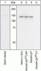
- Peptide Competition. Lysates prepared from CEFs left untransfected (1) or transfected with Src (2-5) were resolved by SDS-PAGE on a 10% polyacrylamide gel and transferred to PVDF. Membranes were blocked with a 5% BSA-TBST buffer for one hour at room temperature and incubated with vinculin (pY822) antibody for one hour at room temperature in 3% BSA-TBST buffer, following prior incubation with: no peptide (1, 2), the non-phosphopeptide corresponding to the immunogen (3), a generic phosphotyrosine-containing peptide (4), or, the phosphopeptide immunogen (5). After washing, membranes were incubated with goat F (ab')2 anti-rabbit IgG HRP conjugate in 3% BSA-TBST buffer, and bands were detected using the Pierce SuperSignal method. The data show that only the peptide corresponding to vinculin (pY822) blocks the antibody signal, thereby demonstrating the specificity of the antibody. The data also show that vinculin is highly phosphorylated in the presence of activated Src.

Supportive validation
- Submitted by
- LSBio (provider)
- Main image

- Experimental details
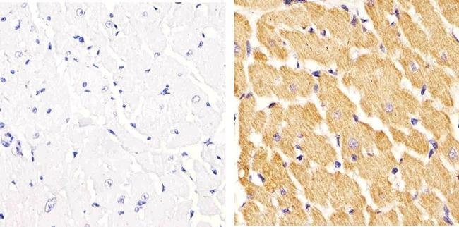
- Immunohistochemistry analysis of Phospho-Vinculin (pTyr822) showing staining in the cytoplasm of paraffin-embedded rat heart tissue (right) compared to a negative control without primary antibody (left). To expose target proteins, antigen retrieval was performed using 10mM sodium citrate (pH 6.0), microwaved for 8-15 min. Following antigen retrieval, tissues were blocked in 3% H2O2-methanol for 15 min at room temperature, washed with ddH2O and PBS, and then probed with a Phospho-Vinculin (pTyr822) polyclonal antibody diluted in 3% BSA-PBS at a dilution of 1:20 overnight at 4°C in a humidified chamber. Tissues were washed extensively in PBST and detection was performed using an HRP-conjugated secondary antibody followed by colorimetric detection using a DAB kit. Tissues were counterstained with hematoxylin and dehydrated with ethanol and xylene to prep for mounting.
- Submitted by
- LSBio (provider)
- Main image

- Experimental details
- Immunohistochemistry analysis of Phospho-Vinculin (pTyr822) showing staining in the cytoplasm of paraffin-embedded mouse heart tissue (right) compared to a negative control without primary antibody (left). To expose target proteins, antigen retrieval was performed using 10mM sodium citrate (pH 6.0), microwaved for 8-15 min. Following antigen retrieval, tissues were blocked in 3% H2O2-methanol for 15 min at room temperature, washed with ddH2O and PBS, and then probed with a Phospho-Vinculin (pTyr822) polyclonal antibody diluted in 3% BSA-PBS at a dilution of 1:20 overnight at 4°C in a humidified chamber. Tissues were washed extensively in PBST and detection was performed using an HRP-conjugated secondary antibody followed by colorimetric detection using a DAB kit. Tissues were counterstained with hematoxylin and dehydrated with ethanol and xylene to prep for mounting.
