Explore
 Validate
 Learn
Learn Western blot
Antibody data
- Antibody Data
- Antigen structure
- References [5]
- Comments [0]
- Validations
- Western blot [2]
- Immunocytochemistry [1]
- Flow cytometry [1]
- Other assay [1]
Submit
Validation data
Reference
Comment
Report error
- Product number
- 44-1074G - Provider product page

- Provider
- Invitrogen Antibodies
- Product name
- Phospho-Vinculin (Tyr100) Polyclonal Antibody
- Antibody type
- Polyclonal
- Antigen
- Synthetic peptide
- Reactivity
- Human, Chicken/Avian
- Host
- Rabbit
- Isotype
- IgG
- Vial size
- 100 µL
- Storage
- -20°C
Submitted references Peptide functionalized DNA hydrogel enhances neuroblastoma cell growth and differentiation.
Expression of CEACAM1 or CEACAM5 in AZ-521 cells restores the type IV secretion deficiency for translocation of CagA by Helicobacter pylori.
6-Mercaptopurine augments glucose transport activity in skeletal muscle cells in part via a mechanism dependent upon orphan nuclear receptor NR4A3.
Paxillin-Y118 phosphorylation contributes to the control of Src-induced anchorage-independent growth by FAK and adhesion.
The Helicobacter pylori CagA protein disrupts matrix adhesion of gastric epithelial cells by dephosphorylation of vinculin.
Hivare P, Gangrade A, Swarup G, Bhavsar K, Singh A, Gupta R, Thareja P, Gupta S, Bhatia D
Nanoscale 2022 Jun 23;14(24):8611-8620
Nanoscale 2022 Jun 23;14(24):8611-8620
Expression of CEACAM1 or CEACAM5 in AZ-521 cells restores the type IV secretion deficiency for translocation of CagA by Helicobacter pylori.
Tegtmeyer N, Harrer A, Schmitt V, Singer BB, Backert S
Cellular microbiology 2019 Jan;21(1):e12965
Cellular microbiology 2019 Jan;21(1):e12965
6-Mercaptopurine augments glucose transport activity in skeletal muscle cells in part via a mechanism dependent upon orphan nuclear receptor NR4A3.
Liu Q, Zhu X, Xu L, Fu Y, Garvey WT
American journal of physiology. Endocrinology and metabolism 2013 Nov 1;305(9):E1081-92
American journal of physiology. Endocrinology and metabolism 2013 Nov 1;305(9):E1081-92
Paxillin-Y118 phosphorylation contributes to the control of Src-induced anchorage-independent growth by FAK and adhesion.
Sachdev S, Bu Y, Gelman IH
BMC cancer 2009 Jan 12;9:12
BMC cancer 2009 Jan 12;9:12
The Helicobacter pylori CagA protein disrupts matrix adhesion of gastric epithelial cells by dephosphorylation of vinculin.
Moese S, Selbach M, Brinkmann V, Karlas A, Haimovich B, Backert S, Meyer TF
Cellular microbiology 2007 May;9(5):1148-61
Cellular microbiology 2007 May;9(5):1148-61
No comments: Submit comment
Supportive validation
- Submitted by
- Invitrogen Antibodies (provider)
- Main image
- Experimental details
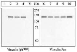
- Peptide Competition: Lysates were prepared from COS cells co-transfected with activated Src and His-tagged chicken vinculin cDNA which were either untreated (1 and 6) or treated with vanadate for 24 hr (2-5 and 7-10).

- Submitted by
- Invitrogen Antibodies (provider)
- Main image

- Experimental details
- Western blot analysis was performed on whole cell extracts (30 µg lysate) of HeLa (Lane 1) and HeLa treated with 100 ng/mL of LPS for 20 mins (Lane 2). The blots were probed with Anti-Phospho-Vinculin pTyr100 Rabbit Polyclonal Antibody (Product # 44-1074G, 1:1000 dilution) and detected by chemiluminescence Goat Anti-Rabbit IgG (H+L) Secondary Antibody, HRP conjugate (Product # G-21234, 1:5000 dilution). Both 116 kDa and 124 kDa bands (isoforms) were observed in HeLa and on treatment with LPS, only a 124 kDa band corresponding to Phospho-Vinculin pTyr100 was observed.Known quantity of protein samples were electrophoresed using Novex® NuPAGE® 4-12 % Bis-Tris gel (Product # NP0341BOX), XCell SureLock™ Electrophoresis System (Product # EI0002) and Novex® Sharp Pre-Stained Protein Standard (Product # LC5800). Resolved proteins were then transferred onto a nitrocellulose membrane by wet transfer. The membrane was probed with the relevant primary and secondary Antibody following blocking with 5 % skimmed milk. Chemiluminescent detection was performed using Pierce™ ECL Western Blotting Substrate (Product # 32106).

Supportive validation
- Submitted by
- Invitrogen Antibodies (provider)
- Main image
- Experimental details
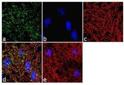
- Immunofluorescence analysis of Phospho-Vinculin pTyr100 was performed using 70% confluent log phase HeLa cells. The cells were fixed with 4% paraformaldehyde for 10 minutes, permeabilized with 0.1% Triton™ X-100 for 10 minutes, and blocked with 2% BSA for 1 hour at room temperature. The cells were labeled with Phospho-Vinculin pTyr100 Rabbit Polyclonal Antibody (Product # 44-1074G) at 1:250 dilution in 0.1% BSA and incubated for 3 hours at room temperature and then labeled with Goat anti-Rabbit IgG (H+L) Superclonal™ Secondary Antibody, Alexa Fluor® 488 conjugate (Product # A27034) a dilution of 1:2000 for 45 minutes at room temperature (Panel a: green). Nuclei (Panel b: blue) were stained with SlowFade® Gold Antifade Mountant with DAPI (Product # S36938). F-actin (Panel c: red) was stained with Rhodamine Phalloidin (Product # R415, 1:300). Panel d represents the merged image showing punctate membranous localization. Panel e shows the no primary antibody control. The images were captured at 60X magnification.

Supportive validation
- Submitted by
- Invitrogen Antibodies (provider)
- Main image
- Experimental details
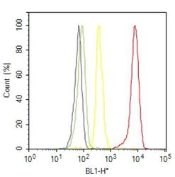
- Flow cytometry analysis of Phospho-Vinculin [pTyr100] was done on K-562 cells treated with Vanadate (100uM, 1 hour). Cells were fixed with 70% ethanol for 10 minutes, permeabilized with 0.25% Triton™ X-100 for 20 minutes, and blocked with 5% BSA for 30 minutes at room temperature. Cells were labeled with Phospho-Vinculin [pTyr100] Rabbit Polyclonal Antibody (441074G, red histogram) or with rabbit isotype control (yellow histogram) at 3-5 ug/million cells in 2.5% BSA. After incubation at room temperature for 2 hours, the cells were labeled with Alexa Fluor® 488 Goat Anti-Rabbit Secondary Antibody (A11008) at a dilution of 1:400 for 30 minutes at room temperature. The representative 10,000 cells were acquired and analyzed for each sample using an Attune® Acoustic Focusing Cytometer. The purple histogram represents unstained control cells and the green histogram represents no-primary-antibody control.

Supportive validation
- Submitted by
- Invitrogen Antibodies (provider)
- Main image
- Experimental details
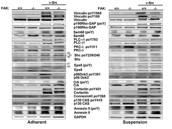
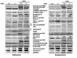
- Figure 2 FAK and adhesion modulate v-Src-induced phosphorylation of various Src substrates . (A) Lysates from adherent cultures of FAK+/+[puro], FAK-/-[puro], FAK+/+[v-Src] and FAK-/-[v-Src] cells were probed either directly by IB for specific phosphorylated form(s) of the Src substrate proteins, total substrate protein levels or GAPDH, or probed for total phosphorylated protein by immunoprecipitating with substrate-specific Ab followed by IB for phosphotyrosine using MAb4G10. (B) Same IB or IP/IB analysis as in panel A using lysates of suspension cultures. Each of these blots is typical of at least duplicate independent experiments.