Explore
 Validate
 Learn
Learn Immunocytochemistry
 Immunohistochemistry
Antibody data
- Antibody Data
- Antigen structure
- References [5]
- Comments [0]
- Validations
- Immunocytochemistry [2]
- Other assay [4]
Submit
Validation data
Reference
Comment
Report error
- Product number
- 53-9777-80 - Provider product page

- Provider
- Invitrogen Antibodies
- Product name
- Vinculin Monoclonal Antibody (7F9), Alexa Fluor™ 488, eBioscience™
- Antibody type
- Monoclonal
- Antigen
- Other
- Description
- Description: The monoclonal antibody 7F9 (VIIF9) recognizes human, mouse, rat, and avian vinculin and its alternatively spliced isoform, metavinculin. Vinculin is a cytoskeletal protein associated with cell-cell and cell-matrix junctions. Vinculin is involved in the anchoring of F-actin to the membrane and the regulation of E-cadherin expression. Vinculin binds to talin, paxillin, and alpha-actinin. Disregulation of vinculin alters cell adhesion, migration, and growth, which promotes cancer invasion and metastasis. Applications Reported: This 7F9 antibody has been reported for use in immunohistochemical staining of frozen tissue sections, immunohistochemical staining of formalin-fixed paraffin embedded tissue sections, microscopy, and immunocytochemistry. Applications Tested: This 7F9 antibody has been tested by immunocytochemistry of formaldehyde-fixed and permeabilized cells and can be used at less than or equal to 20 µg/mL. It is recommended that the antibody be carefully titrated for optimal performance in the assay of interest. Excitation: 488 nm; Emission: 519 nm; Laser: Blue Laser. Filtration: 0.2 µm post-manufacturing filtered.
- Reactivity
- Human, Mouse, Rat
- Host
- Mouse
- Conjugate
- Green dye
- Isotype
- IgG
- Antibody clone number
- 7F9
- Vial size
- 25 µg
- Concentration
- 0.5 mg/mL
- Storage
- 4° C, store in dark, DO NOT FREEZE!
Submitted references ChipCytometry for multiplexed detection of protein and mRNA markers on human FFPE tissue samples.
Miro1-mediated mitochondrial positioning supports subcellular redox status.
Inhibiting IRE1α-endonuclease activity decreases tumor burden in a mouse model for hepatocellular carcinoma.
A MST1-FOXO1 cascade establishes endothelial tip cell polarity and facilitates sprouting angiogenesis.
Substrate properties modulate cell membrane roughness by way of actin filaments.
Jarosch S, Köhlen J, Wagner S, D'Ippolito E, Busch DH
STAR protocols 2022 Jun 17;3(2):101374
STAR protocols 2022 Jun 17;3(2):101374
Miro1-mediated mitochondrial positioning supports subcellular redox status.
Alshaabi H, Shannon N, Gravelle R, Milczarek S, Messier T, Cunniff B
Redox biology 2021 Jan;38:101818
Redox biology 2021 Jan;38:101818
Inhibiting IRE1α-endonuclease activity decreases tumor burden in a mouse model for hepatocellular carcinoma.
Pavlović N, Calitz C, Thanapirom K, Mazza G, Rombouts K, Gerwins P, Heindryckx F
eLife 2020 Oct 26;9
eLife 2020 Oct 26;9
A MST1-FOXO1 cascade establishes endothelial tip cell polarity and facilitates sprouting angiogenesis.
Kim YH, Choi J, Yang MJ, Hong SP, Lee CK, Kubota Y, Lim DS, Koh GY
Nature communications 2019 Feb 19;10(1):838
Nature communications 2019 Feb 19;10(1):838
Substrate properties modulate cell membrane roughness by way of actin filaments.
Chang CH, Lee HH, Lee CH
Scientific reports 2017 Aug 22;7(1):9068
Scientific reports 2017 Aug 22;7(1):9068
No comments: Submit comment
Supportive validation
- Submitted by
- Invitrogen Antibodies (provider)
- Main image
- Experimental details
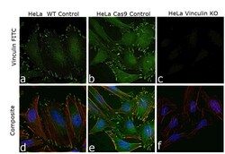
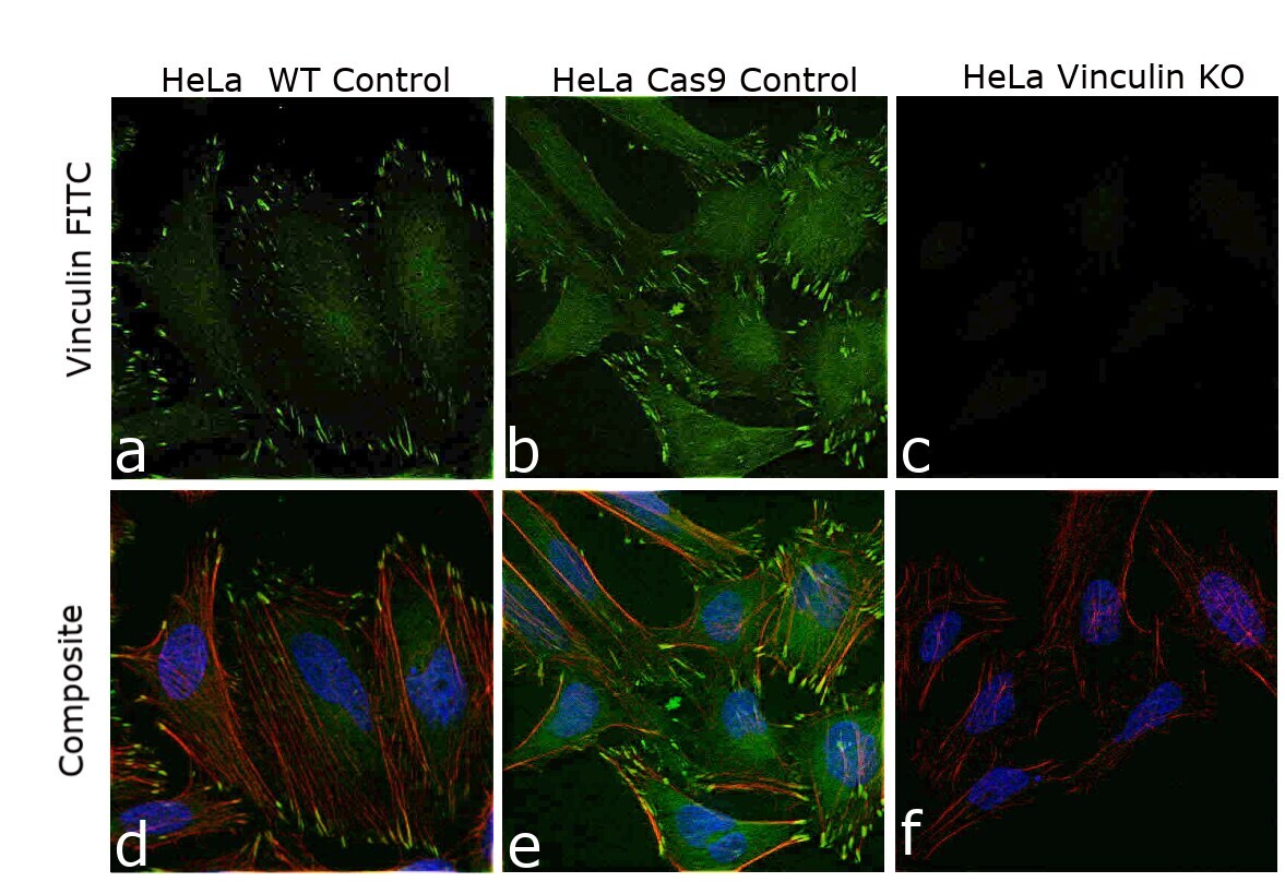
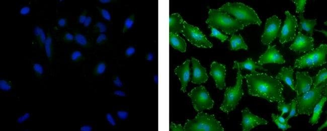
- Knockout of Vinculin was achieved by CRISPR-Cas9 genome editing using LentiArray™ Lentiviral sgRNA (Product # A32042, Assay ID CRISPR948202_LV) and LentiArray Cas9 Lentivirus (Product # A32064). Immunofluorescence analysis was performed on HeLa wild type cells (panel a,d), HeLa Cas9 control cells (panel b,e) and HeLa Vinculin KO cells (panel c, f). Cells were fixed, permeabilized, and labeled with Vinculin Monoclonal Antibody (7F9), Alexa Fluor™ 488, eBioscience™ (Product # 53-9777-82, 1:100 dilution), followed by Donkey anti-Mouse IgG (H+L) Highly Cross-Adsorbed Secondary Antibody, Alexa Fluor™ Plus 488 (Product#A32766, 1:2000 dilution). Nuclei (blue) were stained using ProLong™ Diamond Antifade Mountant with DAPI (Product # P36962), and Rhodamine Phalloidin (Product # R415, 1:300) was used for cytoskeletal F-actin (red) staining. Loss of signal (panel c,f) upon CRISPR mediated knockout (KO) confirms that antibody is specific to Vinculin (green). The images were captured at 60X magnification.
- Conjugate
- Green dye
- Submitted by
- Invitrogen Antibodies (provider)
- Main image
- Experimental details
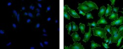
- Immunocytochemistry of formaldehyde-fixed and permeabilized HeLa cells using 20 µg/mL of Mouse IgG1 K Isotype Control Alexa Fluor® 488 (Product # 53-4714-42) (left) or 20 µg/mL of Anti-Vinculin Alexa Fluor® 488 (right). Nuclei are stained with DAPI.
- Conjugate
- Green dye
Supportive validation
- Submitted by
- Invitrogen Antibodies (provider)
- Main image
- Experimental details
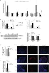
- Figure 2. Increased expression of ER-stress markers in mice with HCC. ( A ) mRNA expression of ER-stress markers Edem1, Ero1b, Grp94, Herp, Atf4, Eif2ak3, Ddit3 , and Hspa5 in liver tissue from healthy mice; and tumor tissue and surrounding non-tumoral tissue from mice with DEN-induced HCC. ( B ) Hspa5- mRNA and ( C ) protein expression of BIP in murine liver tissue. ( D ) Ratio of spliced to unspliced XBP1 in liver tissue from healthy mice; and tumor tissue and surrounding non-tumoral tissue from mice with DEN-induced HCC, treated with 4mu8C. ( E ) Representative western blot image of spliced and unspliced XBP1 protein and vinculin in healthy liver, DEN-induced HCC and DEN-induced HCC treated with 4mu8C. ( F ) quantification of spliced and unspliced XBP1, normalized to total vinculin levels. ( G ) Ratio of spliced to unspliced XBP1 protein levels. ( H ) Representative images and ( I ) quantification of liver tissue sections stained with antibodies against spliced XBP1. p-Values were calculated via the Student''s T-test with five biological replicates per group. Scale bars = 120 mum. Figure 2-figure supplement 1. Activation of the unfolded protein response is mainly located in the stroma of mice with HCC. Liver tissue from mice with DEN-induced HCC, stained with alphaSMA-antibodies and co-stained with antibodies against ( A ) spliced XBP1, ( B ) total XBP1, ( C ) IRE1alpha ( D ) phopho-IRE1alpha, and ( E ) BIP. Scale bars = 50 mum. Figure 2-figure supplement 2. Expression of
- Conjugate
- Green dye
- Submitted by
- Invitrogen Antibodies (provider)
- Main image
- Experimental details
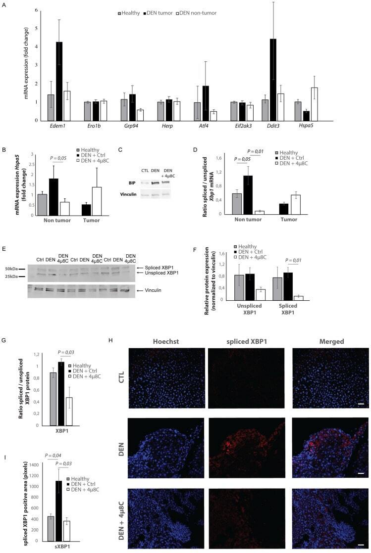
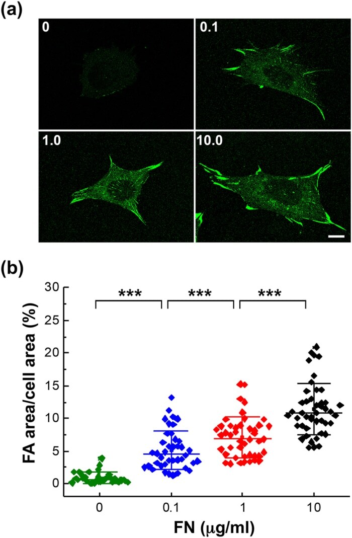
- Figure 2 FA area is positively correlated with the FN concentration on substrate surface. ( a ) MEFs were seeded on the polymer coverslip-bottom mu-dishes coated with ploy-L-lysine followed by 0 to 10 mug/ml FN. After 6 hours, the cells were fixed and stained with anti-vinculin antibody for FA area determination. Scale bar, 10 mum. ( b ) The variation of FA areas in cells responding to various concentrations of FN. Data are expressed as the percentage of the total FA area of each cell relative to the cell area. Values represent mean +- standard deviation ( n = 45 in each condition.). *** P < 0.005 (Student's t-test).
- Conjugate
- Green dye
- Submitted by
- Invitrogen Antibodies (provider)
- Main image

- Experimental details
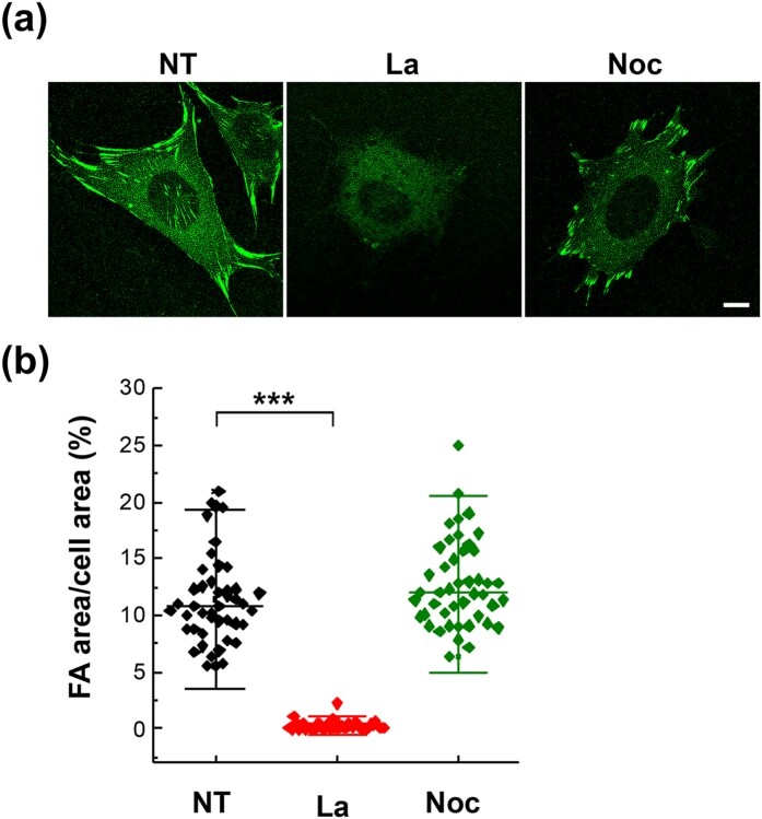
- Figure 4 Actin polymerization is required for FA maturation. ( a ) MEFs were seeded on the 10 mug/ml FN-coated polymer coverslip-bottom mu-dishes and treated with latrunculin (La; 1.0 muM) or nocodazole (Noc; 10 muM) for 30 min followed by immunostaining with anti-vinculin antibody for FA area determination. NT, no treatment. Scale bar, 10 mum. ( b ) The variation of FA areas under various treatments. Data are expressed as the percentage of the total FA area of each cell relative to the cell area. Values represent mean +- standard deviation ( n = 45). *** P < 0.005 (Student's t-test).
- Conjugate
- Green dye
- Submitted by
- Invitrogen Antibodies (provider)
- Main image
- Experimental details
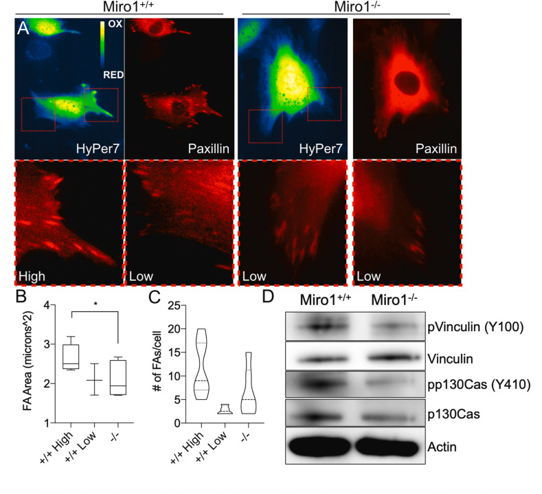
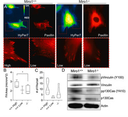
- Fig. 7 Paxillin containing focal adhesion size and abundance correlates with local H 2 O 2 levels. (A) Miro1 +/+ and Miro1 -/- MEFs expressing mCherry-Paxillin and HyPer7. Red-box inserts show mCherry-Paxillin features below at areas of high and low HyPer7 oxidation levels in Miro1 +/+ MEFs and areas of low HyPer7 oxidation in Miro1 -/- MEFs. (B) Quantification of focal adhesion (FA) area (mCherry-Paxillin) at sites of high and low HyPer7 oxidation levels in Miro1 +/+ MEFs and sites of low HyPer7 oxidation in Miro1 -/- MEFs (n = average from 3-5 cells/group, *p < 0.05). (C) Quantification of the number of FAs (mCherry-Paxillin) per cell at sites of high and low HyPer7 oxidation levels in Miro1 +/+ MEFs and sitess of low HyPer7 oxidation in Miro1 -/- MEFs (n = average from 3-5 cells/group). (D) Western blot of reduced phospho-Vinculin (Y100) and phospho-p130Cas (Y410) phosphorylation status in Miro1 -/- MEFs. (For interpretation of the references to colour in this figure legend, the reader is referred to the Web version of this article.) Fig. 7
- Conjugate
- Green dye