Explore
 Validate
 Learn
Learn Western blot
Antibody data
- Antibody Data
- Antigen structure
- References [2]
- Comments [0]
- Validations
- Western blot [3]
- Immunocytochemistry [1]
- Immunohistochemistry [1]
Submit
Validation data
Reference
Comment
Report error
- Product number
- GTX104663 - Provider product page

- Provider
- GeneTex
- Proper citation
- GeneTex Cat#GTX104663, RRID:AB_1241162
- Product name
- PCK2 antibody [C1C2], Internal
- Antibody type
- Polyclonal
- Reactivity
- Human, Mouse, Rat
- Host
- Rabbit
Submitted references Integrating transcriptomics and proteomics to show that tanshinone IIA suppresses cell growth by blocking glucose metabolism in gastric cancer cells.
Quantitative proteomic analysis of human lung tumor xenografts treated with the ectopic ATP synthase inhibitor citreoviridin.
Lin LL, Hsia CR, Hsu CL, Huang HC, Juan HF
BMC genomics 2015 Feb 5;16:41
BMC genomics 2015 Feb 5;16:41
Quantitative proteomic analysis of human lung tumor xenografts treated with the ectopic ATP synthase inhibitor citreoviridin.
Wu YH, Hu CW, Chien CW, Chen YJ, Huang HC, Juan HF
PloS one 2013;8(8):e70642
PloS one 2013;8(8):e70642
No comments: Submit comment
Supportive validation
- Submitted by
- GeneTex (provider)
- Main image
- Experimental details
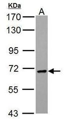
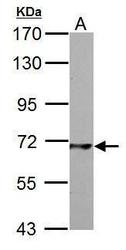
- PCK2 antibody [C1C2], Internal detects PCK2 protein by Western blot analysis.A. 30 ?g HepG2 whole cell lysate/extract7.5 % SDS-PAGEPCK2 antibody [C1C2], Internal (GTX104663) dilution: 1:1000
- Submitted by
- GeneTex (provider)
- Main image
- Experimental details
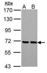
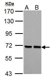
- PCK2 antibody [C1C2], Internal detects PCK2 protein by Western blot analysis.A. 30 ?g Neuro2A whole cell lysate/extractB. 30 ?g GL261 whole cell lysate/extract7.5 % SDS-PAGEPCK2 antibody [C1C2], Internal (GTX104663) dilution: 1:1000
- Submitted by
- GeneTex (provider)
- Main image
- Experimental details
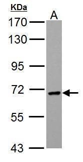
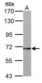
- PCK2 antibody [C1C2], Internal detects PCK2 protein by Western blot analysis.A. 30 ?g PC-12 whole cell lysate/extract7.5 % SDS-PAGEPCK2 antibody [C1C2], Internal (GTX104663) dilution: 1:1000
Supportive validation
- Submitted by
- GeneTex (provider)
- Main image

- Experimental details
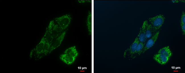
- PCK2 antibody [C1C2], Internal detects PCK2 protein at mitochondria by immunofluorescent analysis.Sample: Hep G2 cells were fixed in 2% paraformaldehyde/culture medium at 37oC for 30 min.Green: PCK2 protein stained by PCK2 antibody [C1C2], Internal (GTX104663) diluted at 1:500.Blue: Hoechst 33342 staining.Scale bar = 10 £gm.
Supportive validation
- Submitted by
- GeneTex (provider)
- Main image

- Experimental details
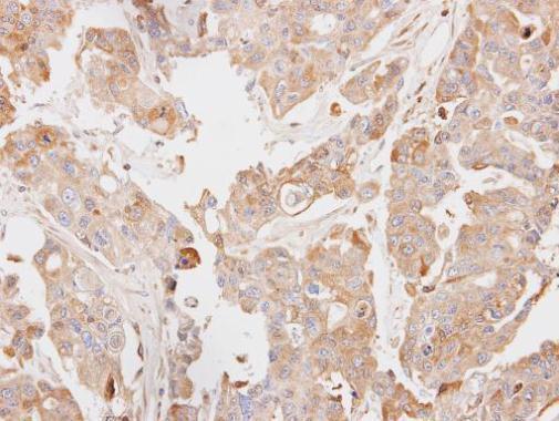
- Immunohistochemical analysis of paraffin-embedded OVCAR3 xenograft, using PCK2(GTX104663) antibody at 1:100 dilution.