Explore
 Validate
 Learn
Learn Western blot
Antibody data
- Antibody Data
- Antigen structure
- References [1]
- Comments [0]
- Validations
- Western blot [3]
- Immunocytochemistry [2]
Submit
Validation data
Reference
Comment
Report error
- Product number
- 702748 - Provider product page

- Provider
- Invitrogen Antibodies
- Product name
- PCK2 Recombinant Rabbit Monoclonal Antibody (16H5L22)
- Antibody type
- Monoclonal
- Antigen
- Synthetic peptide
- Reactivity
- Human
- Host
- Rabbit
- Isotype
- IgG
- Antibody clone number
- 16H5L22
- Vial size
- 100 µg
- Concentration
- 0.5 mg/mL
- Storage
- Store at 4°C short term. For long term storage, store at -20°C, avoiding freeze/thaw cycles.
Submitted references Long non-coding RNA expression profiling following treatment with resveratrol to improve insulin resistance.
Shu L, Hou G, Zhao H, Huang W, Song G, Ma H
Molecular medicine reports 2020 Aug;22(2):1303-1316
Molecular medicine reports 2020 Aug;22(2):1303-1316
No comments: Submit comment
Supportive validation
- Submitted by
- Invitrogen Antibodies (provider)
- Main image

- Experimental details
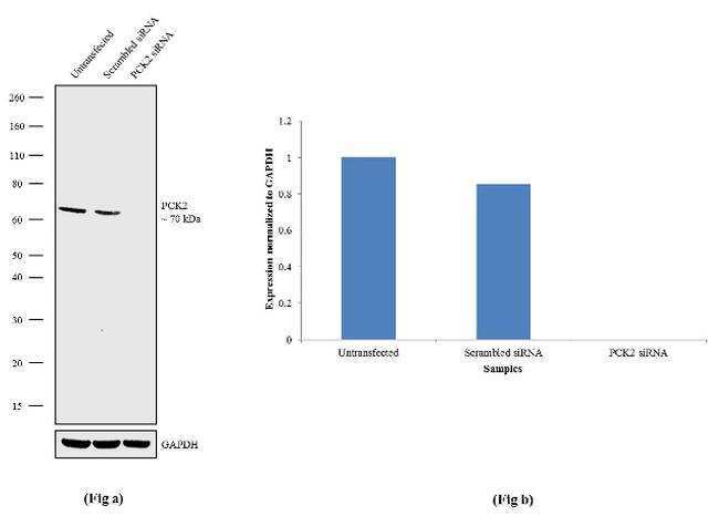
- Knockdown of PCK2 was achieved by transfecting A549 cells with PCK2 specific siRNA (Silencer® select Product # s10126 + s10125). Western blot analysis (Fig a) was performed using Membrane enriched extracts from PCK2 knockdown cells (Lane 3), non-specific scrambled siRNA transfected cells (Lane 2) and untransfected cells (Lane 1). The blots were probed with Anti-PCK2 Recombinant Rabbit Monoclonal Antibody (Product # 702748, 1-3 µg/mL) and Goat anti-Rabbit IgG (H+L) Superclonal™ Secondary Antibody, HRP conjugate (Product # A27036, 0.25 µg/mL, 1:4000 dilution). Densitometric analysis of this Western blot is shown in histogram (Fig b). Loss of signal upon siRNA mediated knock down confirms that antibody is specific to PCK2.
- Submitted by
- Invitrogen Antibodies (provider)
- Main image
- Experimental details
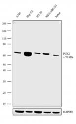
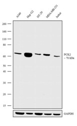
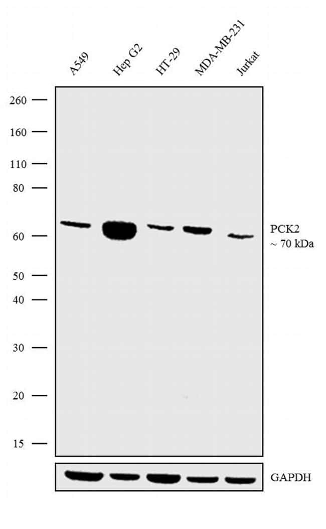
- Western blot analysis was performed on Membrane enriched extracts (30 µg lysate) of A549 (Lane 1), Hep G2 (Lane 2), HT-29 (Lane 3), MDA-MB-231 (Lane 4) and Jurkat (Lane 5). The blots were probed with Anti-PCK2 Recombinant Rabbit Polyclonal Antibody (Product # 702748, 2.5 µg/mL) and detected by chemiluminescence using Goat anti-Rabbit IgG (H+L) Superclonal™ Secondary Antibody, HRP conjugate (Product # A27036, 0.25 µg/mL, 1:4000 dilution). A 70 kDa band corresponding to PCK2 was observed across the cell lines tested. Known quantity of protein samples were electrophoresed using Novex®NuPAGE®4-12% Bis-Tris gel (Product # NP0322BOX), XCell SureLock™ Electrophoresis System (Product # EI0002) and Novex® Sharp Pre-Stained Protein Standard (Product # LC5800). Resolved proteins were then transferred onto a nitrocellulose membrane with iBlot® Dry Blotting System (Product # IB21001). The membrane was probed with the relevant primary and secondary Antibody following blocking with 5% skimmed milk. Chemiluminescent detection was performed using Pierce™ ECL Western blotting Substrate (Product # 32106).

- Submitted by
- Invitrogen Antibodies (provider)
- Main image
- Experimental details
- Western blot analysis was performed on Membrane enriched extracts (30 µg lysate) of A549 (Lane 1), Hep G2 (Lane 2), HT-29 (Lane 3), MDA-MB-231 (Lane 4) and Jurkat (Lane 5). The blots were probed with Anti-PCK2 Recombinant Rabbit Polyclonal Antibody (Product # 702748, 2.5 µg/mL) and detected by chemiluminescence using Goat anti-Rabbit IgG (H+L) Superclonal™ Secondary Antibody, HRP conjugate (Product # A27036, 0.25 µg/mL, 1:4000 dilution). A 70 kDa band corresponding to PCK2 was observed across the cell lines tested. Known quantity of protein samples were electrophoresed using Novex®NuPAGE®4-12% Bis-Tris gel (Product # NP0322BOX), XCell SureLock™ Electrophoresis System (Product # EI0002) and Novex® Sharp Pre-Stained Protein Standard (Product # LC5800). Resolved proteins were then transferred onto a nitrocellulose membrane with iBlot® Dry Blotting System (Product # IB21001). The membrane was probed with the relevant primary and secondary Antibody following blocking with 5% skimmed milk. Chemiluminescent detection was performed using Pierce™ ECL Western blotting Substrate (Product # 32106).
Supportive validation
- Submitted by
- Invitrogen Antibodies (provider)
- Main image

- Experimental details
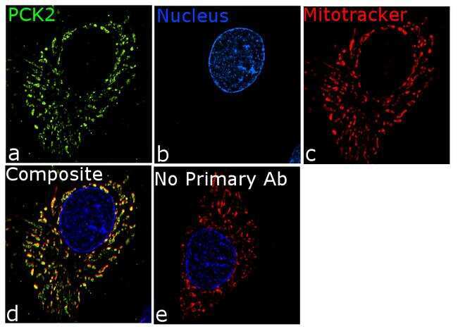
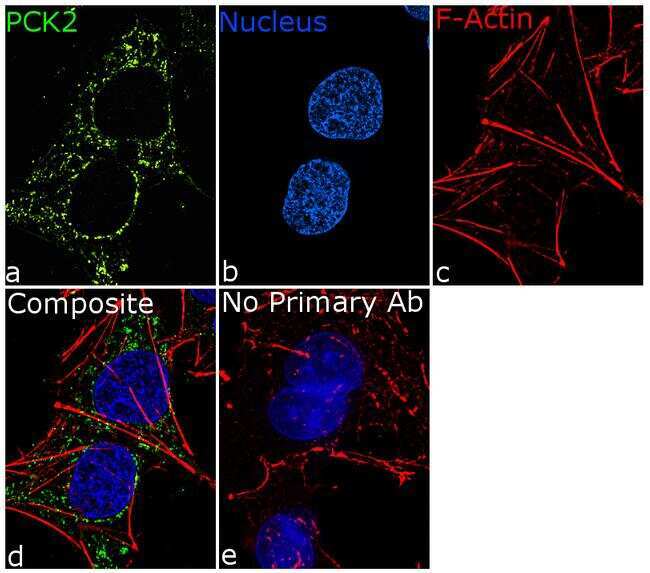
- For immunofluorescence analysis, HepG2 cells were fixed and permeabilized for detection of endogenous PCK2 using Anti- PCK2 Recombinant Rabbit Monoclonal Antibody (Product # 702748, 5 µg/mL) and labeled with Goat anti-Rabbit IgG (H+L) Superclonal™ Secondary Antibody, Alexa Fluor® 488 conjugate (Product # A27034, 1:2000). Panel a) shows representative cells that were stained for detection and localization of PCK2 protein (green), Panel b) is stained for nuclei (blue) using SlowFade® Gold Antifade Mountant with DAPI (Product # S36938). Panel c) represents cytoskeletal F-actin staining using Rhodamine Phalloidin (Product # R415, 1:300). Panel d) is a composite image of Panels a, b and c clearly demonstrating Mitochondrial localization of PCK2. Panel e) represents control cells with no primary antibody to assess background. The images were captured at 60X magnification.
- Submitted by
- Invitrogen Antibodies (provider)
- Main image
- Experimental details
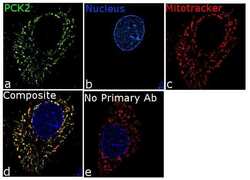
- For immunofluorescence analysis, HepG2 cells were fixed and permeabilized for detection of endogenous PCK2 using Anti- PCK2 Recombinant Rabbit Monoclonal Antibody (Product # 702748, 5 µg/mL) and labeled with Goat anti-Rabbit IgG (H+L) Superclonal™ Secondary Antibody, Alexa Fluor® 488 conjugate (Product # A27034, 1:2000). Panel a) shows representative cells that were stained for detection and localization of PCK2 protein (green), Panel b) is stained for nuclei (blue) using SlowFade® Gold Antifade Mountant with DAPI (Product # S36938). Panel c) represents mitochondrial staining using MitoTracker® Red CMXRos (Product # M7512). Panel d) is a composite image of Panels a, b and c clearly demonstrating co-localization of PCK2 with mitotracker. Panel e) represents control cells with no primary antibody to assess background. The images were captured at 60X magnification.