Explore
 Validate
 Learn
Learn11128-1-AP
antibody from Invitrogen Antibodies
Targeting: MYH9
DFNA17, EPSTS, FTNS, MHA, NMHC-II-A, NMMHCA
 Western blot Immunocytochemistry
 Immunoprecipitation Immunohistochemistry Flow cytometry Other assay
Antibody data
- Antibody Data
- Antigen structure
- References [0]
- Comments [0]
- Validations
- Western blot [6]
- Immunocytochemistry [2]
- Immunohistochemistry [3]
- Flow cytometry [1]
- Other assay [1]
Submit
Validation data
Reference
Comment
Report error
- Product number
- 11128-1-AP - Provider product page

- Provider
- Invitrogen Antibodies
- Product name
- MYH9 Polyclonal Antibody
- Antibody type
- Polyclonal
- Antigen
- Other
- Reactivity
- Human, Mouse, Rat
- Host
- Rabbit
- Isotype
- IgG
- Vial size
- 150 µL
- Concentration
- 0.19 mg/mL
- Storage
- -20°C
No comments: Submit comment
Supportive validation
- Submitted by
- Invitrogen Antibodies (provider)
- Main image
- Experimental details
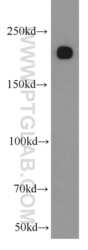
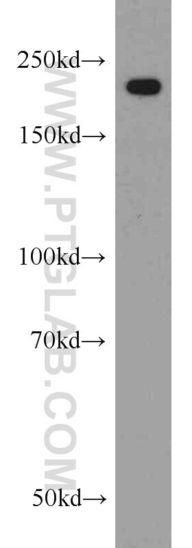
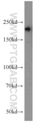
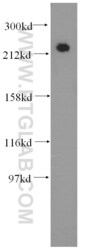
- Mouse kidney tissue were subjected to SDS PAGE followed by western blot with 11128-1-AP (MYH9 antibody) at dilution of 1:1000 incubated at room temperature for 1.5 hours.

- Submitted by
- Invitrogen Antibodies (provider)
- Main image

- Experimental details
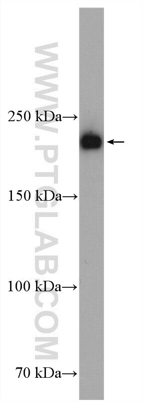
- Mouse brain tissue were subjected to SDS PAGE followed by western blot with 11128-1-AP (MYH9 antibody) at dilution of 1:1000 incubated at room temperature for 1.5 hours.
- Submitted by
- Invitrogen Antibodies (provider)
- Main image
- Experimental details
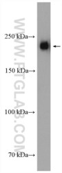
- Mouse colon tissue were subjected to SDS PAGE followed by western blot with 11128-1-AP (MYH9 antibody) at dilution of 1:1000 incubated at room temperature for 1.5 hours.

- Submitted by
- Invitrogen Antibodies (provider)
- Main image
- Experimental details
- Human brain tissue were subjected to SDS PAGE followed by western blot with 11128-1-AP (MYH9 antibody) at dilution of 1:3000 incubated at room temperature for 1.5 hours.

- Submitted by
- Invitrogen Antibodies (provider)
- Main image

- Experimental details
- A549 cells were subjected to SDS PAGE followed by western blot with 11128-1-AP (MYH9 antibody) at dilution of 1:3000 incubated at room temperature for 1.5 hours.
- Submitted by
- Invitrogen Antibodies (provider)
- Main image
- Experimental details
- U2OS cells were subjected to SDS PAGE followed by western blot with 11128-1-AP (MYH9 antibody) at dilution of 1:20000 incubated at room temperature for 1.5 hours.

Supportive validation
- Submitted by
- Invitrogen Antibodies (provider)
- Main image
- Experimental details
- Immunofluorescent analysis of ( -20°C Ethanol ) fixed HepG2 cells using 11128-1-AP (MYH9 antibody) at dilution of 1:50 and Alexa Fluor 488-conjugated AffiniPure Goat Anti-Rabbit IGG (H+L).

- Submitted by
- Invitrogen Antibodies (provider)
- Main image
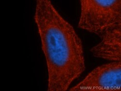
- Experimental details
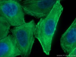
- Immunofluorescent analysis of HepG2 cells, using MYH9 antibody 11128-1-AP at 1:50 dilution and Rhodamine-labeled goat anti-rabbit IgG (red). Blue pseudocolor = DAPI (fluorescent DNA dye).

Supportive validation
- Submitted by
- Invitrogen Antibodies (provider)
- Main image

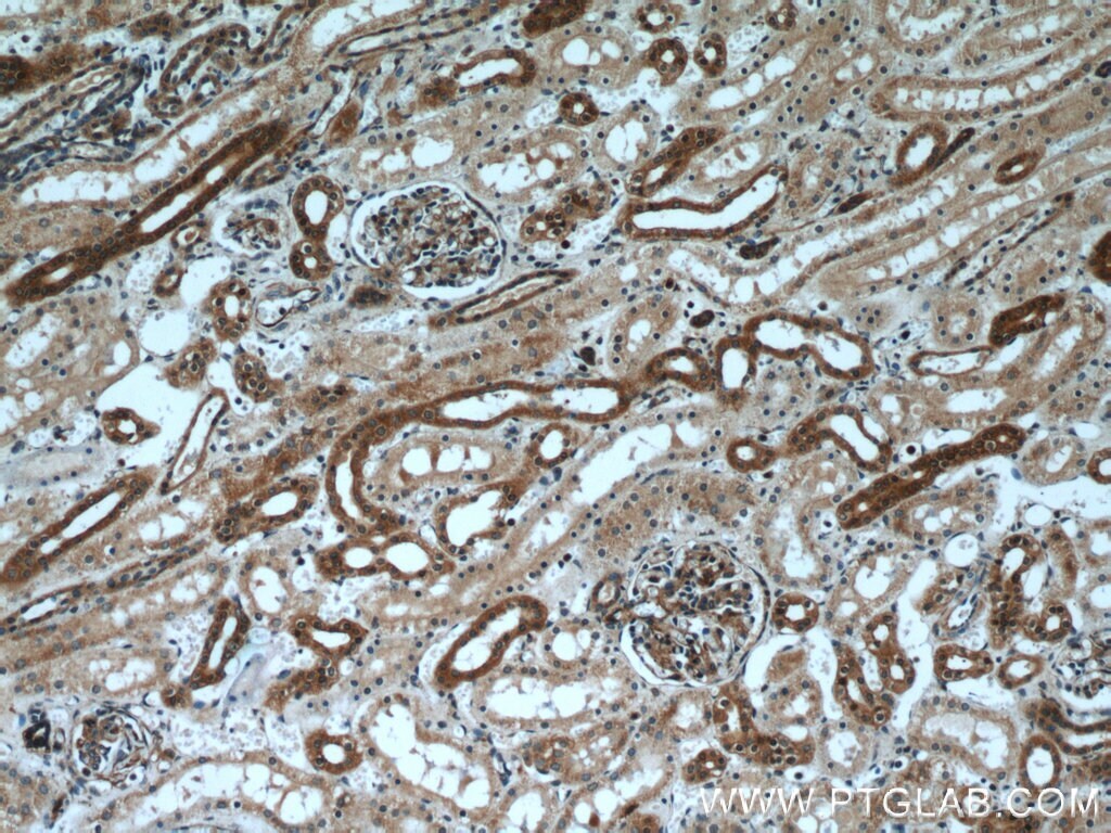
- Experimental details
- Immunohistochemistry of paraffin-embedded human kidney using 11128-1-AP (MYH9 antibody) at dilution of 1:50 (under 10x lens).
- Submitted by
- Invitrogen Antibodies (provider)
- Main image

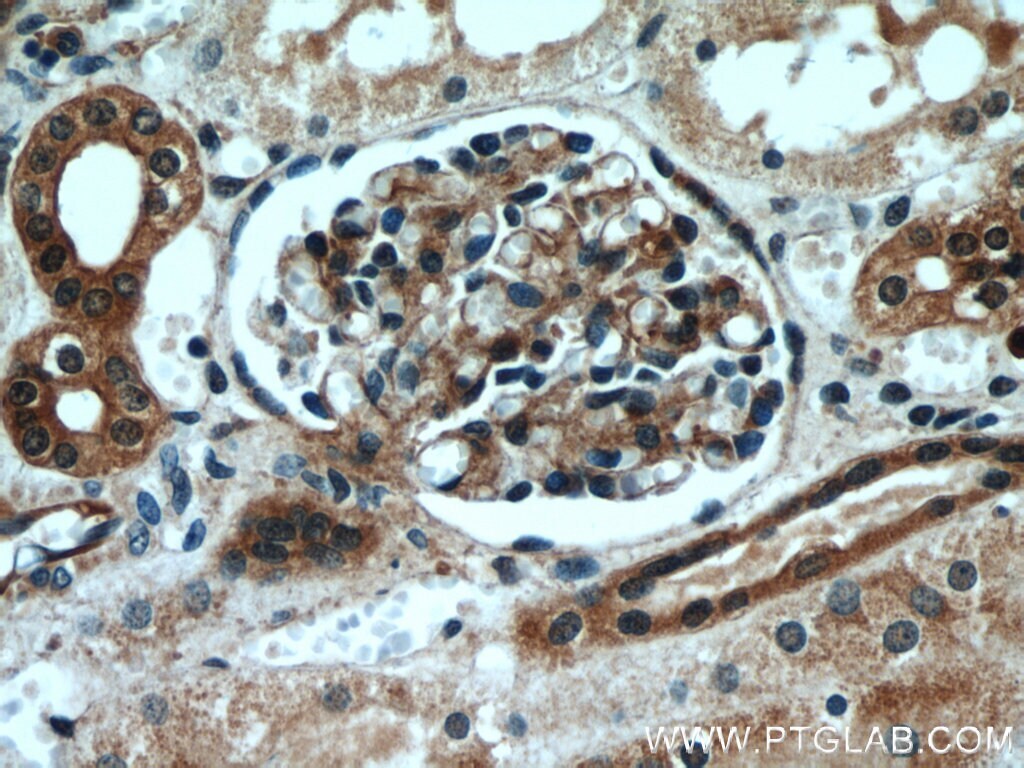
- Experimental details
- Immunohistochemistry of paraffin-embedded human kidney using 11128-1-AP (MYH9 antibody) at dilution of 1:50 (under 40x lens).
- Submitted by
- Invitrogen Antibodies (provider)
- Main image
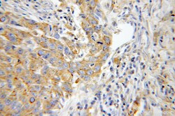
- Experimental details
- Immunohistochemistry of paraffin-embedded human lung cancer using 11128-1-AP (MYH9 antibody) at dilution of 1:100 (under 40x lens).

Supportive validation
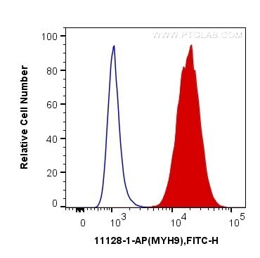
- Submitted by
- Invitrogen Antibodies (provider)
- Main image
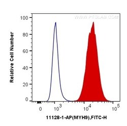
- Experimental details
- 1X10^6 HepG2 cells were intracellularly stained with 0.2 µg Anti-Human MYH9 (Product # 11128-1-AP) and Fluorescein (FITC)-conjugated Affinipure Goat Anti-Rabbit IgG(H+L) at dilution 1:200 (red), or 0.2 µg Control Antibody. Cells were fixed with 90% MeOH .
Supportive validation
- Submitted by
- Invitrogen Antibodies (provider)
- Main image

- Experimental details
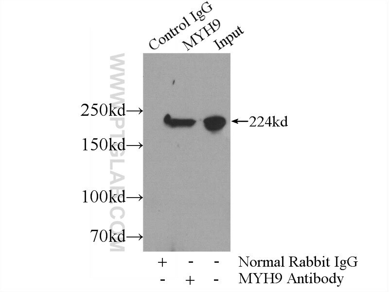
- IP result of anti-MYH9 (IP:11128-1-AP, 5ug; Detection:11128-1-AP 1:1000) with mouse brain tissue lysate 4000ug.