Explore
 Validate
 Learn
Learn Western blot
 Immunohistochemistry
Antibody data
- Antibody Data
- Antigen structure
- References [3]
- Comments [0]
- Validations
- Immunohistochemistry [1]
Submit
Validation data
Reference
Comment
Report error
- Product number
- A00110-1 - Provider product page

- Provider
- Boster Biological Technology
- Product name
- Anti-SP1 Antibody Picoband™
- Antibody type
- Polyclonal
- Description
- Rabbit IgG polyclonal antibody for SP1 detection. Tested with WB, IHC-P, ICC/IF, Direct ELISA in Human;Mouse;Rat.
- Reactivity
- Human, Mouse, Rat
- Host
- Rabbit
- Vial size
- 100μg/vial
- Concentration
- 0.5-1mg/ml, actual concentration vary by lot. Use suggested dilution ratio to decide dilution procedure.
- Storage
- At -20°C for one year. After reconstitution, at 4°C for one month. It can also be aliquoted and stored frozen at -20°C for a longer time. Avoid repeated freezing and thawing.
- Handling
- Add 0.2ml of distilled water will yield a concentration of 500ug/ml.
Submitted references Epigenetic Regulation of TET1-SP1 During Spermatogonia Self-Renewal and Proliferation.
Chi-miR-4110 promotes granulosa cell apoptosis by targeting Sma- and Mad-related protein 2 (Smad2) in the caprine ovary.
Vascular endothelial growth factor induces multidrug resistance-associated protein 1 overexpression through phosphatidylinositol-3-kinase /protein kinase B signaling pathway and transcription factor specificity protein 1 in BGC823 cell line.
Liu L, Wang J, Wang S, Wang M, Chen Y, Zheng L
Frontiers in physiology 2022;13:843825
Frontiers in physiology 2022;13:843825
Chi-miR-4110 promotes granulosa cell apoptosis by targeting Sma- and Mad-related protein 2 (Smad2) in the caprine ovary.
An X, Song Y, Hou J, Zhang Y, Chen K, Ma H, Zhao X, Li G, Gao K, Wang S, Cao B, Bai Y
PloS one 2017;12(7):e0181162
PloS one 2017;12(7):e0181162
Vascular endothelial growth factor induces multidrug resistance-associated protein 1 overexpression through phosphatidylinositol-3-kinase /protein kinase B signaling pathway and transcription factor specificity protein 1 in BGC823 cell line.
Li J, Wu X, Gong J, Yang J, Leng J, Chen Q, Xu W
Acta biochimica et biophysica Sinica 2013 Aug;45(8):656-63
Acta biochimica et biophysica Sinica 2013 Aug;45(8):656-63
No comments: Submit comment
Supportive validation
- Submitted by
- Boster Biological Technology (provider)
- Main image

- Experimental details
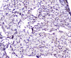
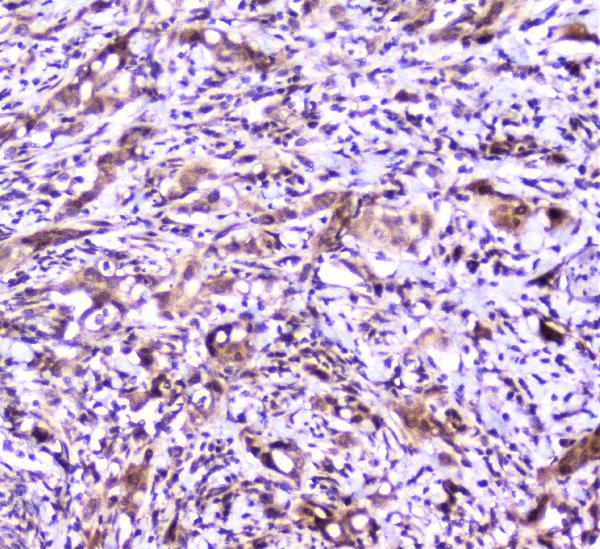
- IHC analysis of SP1 using anti-SP1 antibody (A00110-1).SP1 was detected in paraffin-embedded section of human intestinal cancer tissue. Heat mediated antigen retrieval was performed in citrate buffer (pH6, epitope retrieval solution) for 20 mins. The tissue section was blocked with 10% goat serum. The tissue section was then incubated with 2μg/ml rabbit anti-SP1 Antibody (A00110-1) overnight at 4°C. Biotinylated goat anti-rabbit IgG was used as secondary antibody and incubated for 30 minutes at 37°C. The tissue section was developed using Strepavidin-Biotin-Complex (SABC)(Catalog # SA1022) with DAB as the chromogen.
- Additional image