Explore
 Validate
 Learn
Learn Western blot
 Immunoprecipitation
Antibody data
- Antibody Data
- Antigen structure
- References [4]
- Comments [0]
- Validations
- Western blot [2]
- Immunohistochemistry [1]
Submit
Validation data
Reference
Comment
Report error
- Product number
- NB600-232 - Provider product page

- Provider
- Novus Biologicals
- Proper citation
- Novus Cat#NB600-232, RRID:AB_10000557
- Product name
- Rabbit Polyclonal SP1 Antibody
- Antibody type
- Polyclonal
- Description
- Immunogen affinity purified.
- Reactivity
- Human, Mouse
- Host
- Rabbit
- Isotype
- IgG
- Vial size
- 100 ul
- Concentration
- 1.0 mg/ml
- Storage
- Store at 4C. Do not freeze.
Submitted references HIF2α-Sp1 interaction mediates a deacetylation-dependent FVII-gene activation under hypoxic conditions in ovarian cancer cells.
Regulation of monoamine oxidase A by the SRY gene on the Y chromosome.
Retinoic acid activates monoamine oxidase B promoter in human neuronal cells.
Involvement of the GC-rich sequence and specific proteins (Sp1/Sp3) in the basal transcription activity of neurogranin gene.
Koizume S, Ito S, Miyagi E, Hirahara F, Nakamura Y, Sakuma Y, Osaka H, Takano Y, Ruf W, Miyagi Y
Nucleic acids research 2012 Jul;40(12):5389-401
Nucleic acids research 2012 Jul;40(12):5389-401
Regulation of monoamine oxidase A by the SRY gene on the Y chromosome.
Wu JB, Chen K, Li Y, Lau YF, Shih JC
FASEB journal : official publication of the Federation of American Societies for Experimental Biology 2009 Nov;23(11):4029-38
FASEB journal : official publication of the Federation of American Societies for Experimental Biology 2009 Nov;23(11):4029-38
Retinoic acid activates monoamine oxidase B promoter in human neuronal cells.
Wu JB, Chen K, Ou XM, Shih JC
The Journal of biological chemistry 2009 Jun 19;284(25):16723-35
The Journal of biological chemistry 2009 Jun 19;284(25):16723-35
Involvement of the GC-rich sequence and specific proteins (Sp1/Sp3) in the basal transcription activity of neurogranin gene.
Gui J, Song Y, Han NL, Zhou SF, Sheu FS
Biochemical and biophysical research communications 2006 Jun 23;345(1):124-32
Biochemical and biophysical research communications 2006 Jun 23;345(1):124-32
No comments: Submit comment
Supportive validation
- Submitted by
- Novus Biologicals (provider)
- Main image
- Experimental details
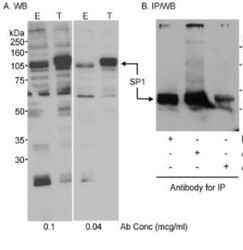
- Western Blot: SP1 Antibody [NB600-232] - A. Whole cell lysate (50 ug for E; 10 ug for T) from HEK 293T cells that were mock transfected (E) or transfected with an Sp1 expression construct (T). B. Whole cell lysate (~700 ug) from HEK 293T cells. A. Affinity purified rabbit anti-Sp1 antibody used at the indicated concentrations. B. SP1 was immunoprecipitated using affinity purified rabbit anti-SP1 antibodies BL937, using each antibody at 3.75 ug/mg lysate. Subsequently, was used at 0.04 ug/mL for WB. Detection by chemiluminescence with an exposure time of 10 seconds (A) or 1 min (B).

- Submitted by
- Novus Biologicals (provider)
- Main image
- Experimental details
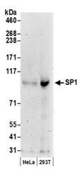
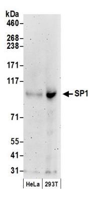
- Western Blot: SP1 Antibody [NB600-232] - Detection of Human SP1 by Western Blot. Whole cell lysate (50 ug) from HeLa and 293T cells prepared using NETN lysis buffer. Affinity purified rabbit anti-SP1 antibody (NB600-232) used for WB at 0.04 ug/mL. Detection by chemiluminescence with an exposure time of 3 minutes.
Supportive validation
- Submitted by
- Novus Biologicals (provider)
- Main image
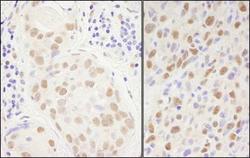
- Experimental details
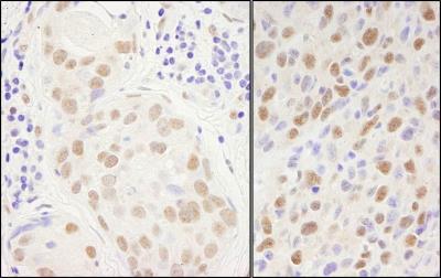
- Immunohistochemistry-Paraffin: SP1 Antibody [NB600-232] - FFPE sections of human breast carcinoma (left) and mouse squamous cell carcinoma (right). Affinity purified rabbit anti-Sp1 used at a dilution of 1:1,000 (1 ug/mL). Detection by DAB.