Explore
 Validate
 Learn
Learn Flow cytometry
Antibody data
- Antibody Data
- Antigen structure
- References [12]
- Comments [0]
- Validations
- Flow cytometry [1]
- Other assay [3]
Submit
Validation data
Reference
Comment
Report error
- Product number
- 12-9320-80 - Provider product page

- Provider
- Invitrogen Antibodies
- Product name
- PLZF Monoclonal Antibody (Mags.21F7), PE, eBioscience™
- Antibody type
- Monoclonal
- Antigen
- Other
- Description
- Description: This Mags.21F7 monoclonal antibody reacts with human and mouse promyelocytic leukemia zinc finger (PLZF), a member of the BTB-POZ family of transcription factors. Expression of this transcriptional repressor in immune cells differs between mice and humans. In mice, PLZF is highly expressed in immature CD1d-resricted NKT2 and NKT1 cells, and a subset of gamma delta (Vg1.1+Vd6.3+) T cells. Studies have also demonstrated expression of PLZF in non-invariant CD1d-restricted T cells, as well as non-CD1d-restricted innate T cells. In humans, PLZF is expressed in NK cells, gamma delta T cells, as well as CD4 and CD8+ T cells. PLZF is also expressed in MR1-specific mucosal-associated invariant T cells, as well as in MHC Class II-restricted T cells that develop via a thymocyte-thymocyte interaction in humans. PLZF exists as a homodimer or in complex with PLZP, and has been shown to be involved in the development of NKT cells, NK cell function, cellular quiescence, and growth suppression. Finally, PLZF has been shown to inhibit gene expression induced by retinoic acid receptor. Applications Reported: This Mags.21F7 antibody has been reported for use in intracellular staining followed by flow cytometric analysis. Applications Tested: This Mags.21F7 antibody has been tested by intracellular staining followed by flow cytometric analysis of mouse lymph node cells using the Foxp3/Transcription Factor Staining Buffer Set (Product # 00-5523-00) and protocol. This can be used at less than or equal to 0.5 µg per test. A test is defined as the amount (µg) of antibody that will stain a cell sample in a final volume of 100 µL. Cell number should be determined empirically but can range from 10^5 to 10^8 cells/test. It is recommended that the antibody be carefully titrated for optimal performance in the assay of interest. Excitation: 488-561 nm; Emission: 578 nm; Laser: Blue Laser, Green Laser, Yellow-Green Laser. Filtration: 0.2 µm post-manufacturing filtered.
- Reactivity
- Human, Mouse
- Host
- Mouse
- Conjugate
- Yellow dye
- Isotype
- IgG
- Antibody clone number
- Mags.21F7
- Vial size
- 25 µg
- Concentration
- 0.2 mg/mL
- Storage
- 4° C, store in dark, DO NOT FREEZE!
Submitted references Loss of Ubiquitin Carboxy-Terminal Hydrolase L1 Impairs Long-Term Differentiation Competence and Metabolic Regulation in Murine Spermatogonial Stem Cells.
Thymic iNKT single cell analyses unmask the common developmental program of mouse innate T cells.
Transnuclear mice reveal Peyer's patch iNKT cells that regulate B-cell class switching to IgG1.
Commensal Microbiota Promote Lung Cancer Development via γδ T Cells.
In Vitro Cytotoxicity of Folate-Silica-Gold Nanorods on Mouse Acute Lymphoblastic Leukemia and Spermatogonial Cells.
Invariant natural killer T-cell development and function with loss of microRNA-155.
Reactive Oxygen Species Regulate the Inflammatory Function of NKT Cells through Promyelocytic Leukemia Zinc Finger.
MR1-restricted mucosal-associated invariant T (MAIT) cells respond to mycobacterial vaccination and infection in nonhuman primates.
Systemic Human ILC Precursors Provide a Substrate for Tissue ILC Differentiation.
The Hypothesis of the Human iNKT/Innate CD8(+) T-Cell Axis Applied to Cancer: Evidence for a Deficiency in Chronic Myeloid Leukemia.
Id2 regulates hyporesponsive invariant natural killer T cells.
E and Id proteins influence invariant NKT cell sublineage differentiation and proliferation.
Alpaugh WF, Voigt AL, Dardari R, Su L, Al Khatib I, Shin W, Goldsmith TM, Coyle KM, Tang LA, Shutt TE, Klein C, Biernaskie J, Dobrinski I
Cells 2021 Aug 31;10(9)
Cells 2021 Aug 31;10(9)
Thymic iNKT single cell analyses unmask the common developmental program of mouse innate T cells.
Harsha Krovi S, Zhang J, Michaels-Foster MJ, Brunetti T, Loh L, Scott-Browne J, Gapin L
Nature communications 2020 Dec 7;11(1):6238
Nature communications 2020 Dec 7;11(1):6238
Transnuclear mice reveal Peyer's patch iNKT cells that regulate B-cell class switching to IgG1.
Clancy-Thompson E, Chen GZ, LaMarche NM, Ali LR, Jeong HJ, Crowley SJ, Boelaars K, Brenner MB, Lynch L, Dougan SK
The EMBO journal 2019 Jul 15;38(14):e101260
The EMBO journal 2019 Jul 15;38(14):e101260
Commensal Microbiota Promote Lung Cancer Development via γδ T Cells.
Jin C, Lagoudas GK, Zhao C, Bullman S, Bhutkar A, Hu B, Ameh S, Sandel D, Liang XS, Mazzilli S, Whary MT, Meyerson M, Germain R, Blainey PC, Fox JG, Jacks T
Cell 2019 Feb 21;176(5):998-1013.e16
Cell 2019 Feb 21;176(5):998-1013.e16
In Vitro Cytotoxicity of Folate-Silica-Gold Nanorods on Mouse Acute Lymphoblastic Leukemia and Spermatogonial Cells.
Eslahi N, Shakeri-Zadeh A, Ashtari K, Pirhajati-Mahabadi V, Tohidi Moghadam T, Shabani R, Kamrava K, Madjd Z, Maki C, Asgari HR, Koruji M
Cell journal 2019 Apr;21(1):14-26
Cell journal 2019 Apr;21(1):14-26
Invariant natural killer T-cell development and function with loss of microRNA-155.
Frias AB Jr, Buechel HM, Neupane A, D'Cruz LM
Immunology 2018 Feb;153(2):238-245
Immunology 2018 Feb;153(2):238-245
Reactive Oxygen Species Regulate the Inflammatory Function of NKT Cells through Promyelocytic Leukemia Zinc Finger.
Kim YH, Kumar A, Chang CH, Pyaram K
Journal of immunology (Baltimore, Md. : 1950) 2017 Nov 15;199(10):3478-3487
Journal of immunology (Baltimore, Md. : 1950) 2017 Nov 15;199(10):3478-3487
MR1-restricted mucosal-associated invariant T (MAIT) cells respond to mycobacterial vaccination and infection in nonhuman primates.
Greene JM, Dash P, Roy S, McMurtrey C, Awad W, Reed JS, Hammond KB, Abdulhaqq S, Wu HL, Burwitz BJ, Roth BF, Morrow DW, Ford JC, Xu G, Bae JY, Crank H, Legasse AW, Dang TH, Greenaway HY, Kurniawan M, Gold MC, Harriff MJ, Lewinsohn DA, Park BS, Axthelm MK, Stanton JJ, Hansen SG, Picker LJ, Venturi V, Hildebrand W, Thomas PG, Lewinsohn DM, Adams EJ, Sacha JB
Mucosal immunology 2017 May;10(3):802-813
Mucosal immunology 2017 May;10(3):802-813
Systemic Human ILC Precursors Provide a Substrate for Tissue ILC Differentiation.
Lim AI, Li Y, Lopez-Lastra S, Stadhouders R, Paul F, Casrouge A, Serafini N, Puel A, Bustamante J, Surace L, Masse-Ranson G, David E, Strick-Marchand H, Le Bourhis L, Cocchi R, Topazio D, Graziano P, Muscarella LA, Rogge L, Norel X, Sallenave JM, Allez M, Graf T, Hendriks RW, Casanova JL, Amit I, Yssel H, Di Santo JP
Cell 2017 Mar 9;168(6):1086-1100.e10
Cell 2017 Mar 9;168(6):1086-1100.e10
The Hypothesis of the Human iNKT/Innate CD8(+) T-Cell Axis Applied to Cancer: Evidence for a Deficiency in Chronic Myeloid Leukemia.
Jacomet F, Cayssials E, Barbarin A, Desmier D, Basbous S, Lefèvre L, Levescot A, Robin A, Piccirilli N, Giraud C, Guilhot F, Roy L, Herbelin A, Gombert JM
Frontiers in immunology 2016;7:688
Frontiers in immunology 2016;7:688
Id2 regulates hyporesponsive invariant natural killer T cells.
Stradner MH, Cheung KP, Lasorella A, Goldrath AW, D'Cruz LM
Immunology and cell biology 2016 Aug;94(7):640-5
Immunology and cell biology 2016 Aug;94(7):640-5
E and Id proteins influence invariant NKT cell sublineage differentiation and proliferation.
D'Cruz LM, Stradner MH, Yang CY, Goldrath AW
Journal of immunology (Baltimore, Md. : 1950) 2014 Mar 1;192(5):2227-36
Journal of immunology (Baltimore, Md. : 1950) 2014 Mar 1;192(5):2227-36
No comments: Submit comment
Supportive validation
- Submitted by
- Invitrogen Antibodies (provider)
- Main image
- Experimental details
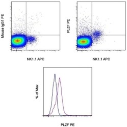
- TOP: C57Bl/6 thymocytes were stained with Anti-Mouse NK1-1 APC (Product # 17-5941-82) and Anti-Mouse TCR beta eFluor 450 (Product # 48-5961-82) followed by intracellular staining with 0.25 µg Mouse IgG1 K Isotype Control PE (Product # 12-4714-81) (left) or Anti-Mouse PLZF PE (right) using the Foxp3 Staining Buffer Set and protocol (Product # 00-5523-00). TCR beta+ cells in the lymphocyte gate were used for analysis. BOTTOM: C57Bl/6 thymocytes were stained with Anti-Mouse NK1-1 APC (Product # 17-5941-82) and Anti-Mouse TCR beta eFluor 450 (Product # 48-5961-82) followed by intracellular staining with 0.25 µg Mouse IgG1 K Isotype Control PE (Product # 12-4714-81) (blue histogram) or Anti-Mouse PLZF PE (purple histogram) using the Foxp3 Staining Buffer Set and protocol (Product # 00-5523-00). NK1-1+TCR beta+ cells in the lymphocyte gate were used for analysis.
- Conjugate
- Yellow dye

Supportive validation
- Submitted by
- Invitrogen Antibodies (provider)
- Main image
- Experimental details
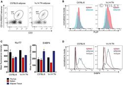
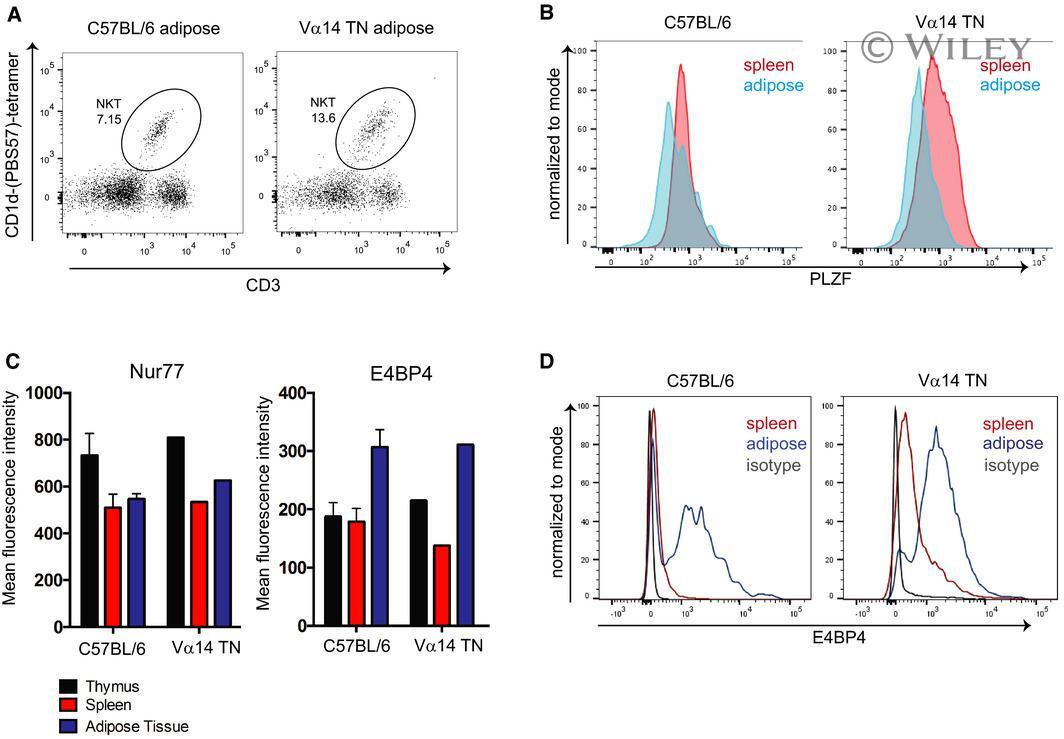
- EV2 Adipose iNKT cells from Valpha14 TN mice are indistinguishable from C57 BL /6-derived adipose iNKT cells Flow cytometry analysis of iNKT cell abundance in white adipose tissue from a Valpha14 TN mouse. Spleen cells and stromal vascular fractions of white adipose tissue from Valpha14 TN or C57BL/6 mice were stained intracellularly with anti-PLZF and analyzed by flow cytometry. Histograms shown are gated on CD1d-(PBS57)-tetramer + CD3 + cells. Thymus, spleen, and adipose tissue were harvested from C57BL/6 mice and Valpha14 TN mice. Cell suspensions were stained with antibodies to CD3, Nur77, E4BP4, and CD1d-(PBS57)-tetramer and analyzed by flow cytometry. Mean fluorescence intensity of Nur77 and E4BP4 staining after gating on iNKT cells is shown. N = 3 per group. Error bars are SEM. Representative histograms of E4BP4 staining are shown. Plots are gated on total CD3 + cells.
- Conjugate
- Yellow dye
- Submitted by
- Invitrogen Antibodies (provider)
- Main image
- Experimental details
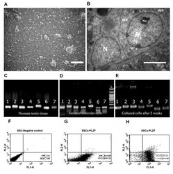
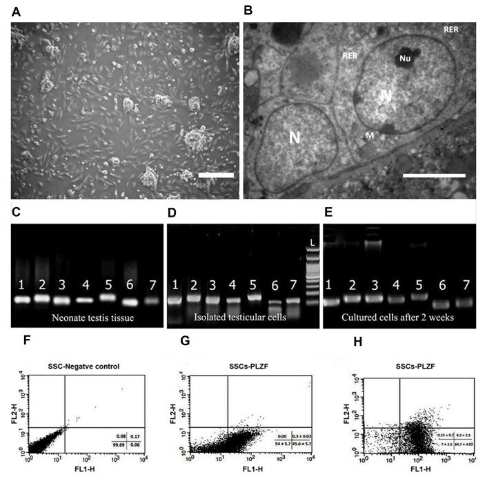
- Fig.1 Spermatogonial cells characterization. A . The morphology of a spermatogonial-derived cluster formed from the culturing of spermatogonial cells after 24 hours (scale bar: 200 um), B . Representative transmission electron micrographs from spermatogonial cells (SSCs) clusters (scale bar: 5 um). The heterochromatin nucleus (N), eccentric small compact and highly reticulated nucleoli (Nu), Rough endoplasmic reticulum (RER) and very high mitochondria (M) were observed in cells. Reverse transcription polymerase chain reaction (RT-PCR) was used to determine the expression of specific spermatogonia and germ cell markers in C . Neonate testis tissue (fresh tissue without enzymatic digestion), D . Cultured cells after the first day and E . Two weeks of culture. 1; Oct4 (129 bp), 2; Itgalpha6 (148 bp), 3; Plzf (137 bp), 4; Gfralpha1 (130 bp), 5; Mvh (Vasa, 149 bp), 6; Itgss1 (115 bp), 7; Gapdh (125 bp). Flow cytometric analysis of spermatogonial cells: F . Spermatogonial negative control, G . The PLZF positive spermatogonial cells at the end of the first week were 45.63 +- 5.71%, and H . At the end of the second week was 84.68 +- 4.02%.
- Conjugate
- Yellow dye
- Submitted by
- Invitrogen Antibodies (provider)
- Main image
- Experimental details
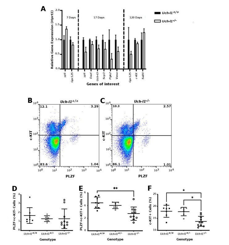
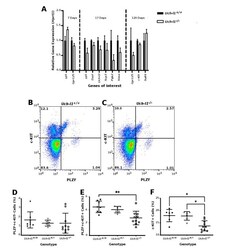
- Figure 3 Analysis of whole Uch-l1 -/- , Uch-l1 +/- and Uch-l1 +/+ testes. ( A ) Quantitative PCR results of significantly differentially expressed genes in 7, 17 and 120-day old of Uch-l1 -/- relative to Uch-l1 +/+ testes ( B , F ) Flow cytometry analysis of PLZF and c-KIT positive cells in Uch-l1 +/+ , Uch-l1 +/- and Uch-l1 -/- testes at 120 days of age. n = 6/genotype. ( B , C ) Representative dot plot of c-KIT and PLZF positive cells in Uch-l1 +/+ and Uch-l1 -/- testes, respectively. ( D ) Percentage of PLZF+ c-KIT- cells in Uch-l1 +/+ , Uch-l1 +/- and Uch-l1 -/- . ( E ) Percentage of PLZF+ c-KIT+ cells in Uch-l1 +/+ , Uch-l1 +/- and Uch-l1 -/- . ( F ) Percentage of c-KIT+ cells in Uch-l1 +/+ , Uch-l1 +/- and Uch-l1 -/- . * indicates significant differences ( p < 0.05), ** represents significant differences p < 0.01.
- Conjugate
- Yellow dye