Explore
 Validate
 Learn
Learn Flow cytometry
Antibody data
- Antibody Data
- Antigen structure
- References [2]
- Comments [0]
- Validations
- Flow cytometry [1]
Submit
Validation data
Reference
Comment
Report error
- Product number
- 25-9322-82 - Provider product page

- Provider
- Invitrogen Antibodies
- Product name
- PLZF Monoclonal Antibody (9E12), PE-Cyanine7, eBioscience™
- Antibody type
- Monoclonal
- Antigen
- Synthetic peptide
- Description
- This 9E12 monoclonal antibody reacts with human and mouse promyelocytic leukemia zinc finger (PLZF), a member of the BTB-POZ family of transcription factors. Expression of this transcriptional repressor in immune cells differs between mice and humans. In mice, PLZF is highly expressed in immature CD1d-resricted NKT2 and NKT1 cells, and a subset of gamma delta (Vg1.1+Vd6.3+) T cells. Studies have also demonstrated expression of PLZF in non-invariant CD1d-restricted T cells, as well as non-CD1d-restricted innate T cells. In humans, PLZF is expressed in NK cells, gamma delta T cells, as well as CD4 and CD8+ T cells. PLZF is also expressed in MR1-specific mucosal-associated invariant T cells, as well as in MHC Class II-restricted T cells that develop via a thymocyte-thymocyte interaction in humans. PLZF is expressed by innate lymphoid cells and its progenitor cells. PLZF exists as a homodimer or in complex with PLZP, and has been shown to be involved in the development of NKT cells, NK cell function, cellular quiescence, growth suppression, and maintenance of homeostatis. Finally, PLZF has been shown to inhibit gene expression induced by retinoic acid receptor.
- Antibody clone number
- 9E12
- Concentration
- 0.2 mg/mL
Submitted references Identification and Isolation of Type II NKT Cell Subsets in Human Blood and Liver.
Thymic iNKT single cell analyses unmask the common developmental program of mouse innate T cells.
Yang Zhou J, Werner JM, Glehr G, Geissler EK, Hutchinson JA, Kronenberg K
Frontiers in immunology 2022;13:898473
Frontiers in immunology 2022;13:898473
Thymic iNKT single cell analyses unmask the common developmental program of mouse innate T cells.
Harsha Krovi S, Zhang J, Michaels-Foster MJ, Brunetti T, Loh L, Scott-Browne J, Gapin L
Nature communications 2020 Dec 7;11(1):6238
Nature communications 2020 Dec 7;11(1):6238
No comments: Submit comment
Supportive validation
- Submitted by
- Invitrogen Antibodies (provider)
- Main image

- Experimental details
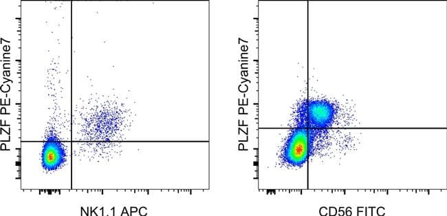
- LEFT: C57BL/6 thymocytes were surface stained with Anti-Mouse NK1.1 APC (Product # 17-5941-63) and Anti-Mouse TCR beta FITC (Product # 11-5961-82) followed by intracellular staining with 0.125 µg of Anti-Human/Mouse PLZF PE-Cyanine7 using the Foxp3/Transcription Factor Staining Buffer Set and protocol (Product # 00-5523-00). Viable, TCR beta+ cells, as determined by Fixable Viability Dye eFluor® 450 (Product # 65-0863-14), were used for analysis. RIGHT: Normal human peripheral blood cells were surface stained with Anti-Human CD56 (NCAM) FITC (Product # 11-0566-42) followed by intracellular staining with 0.125 µg of Anti-Human/Mouse PLZF PE-Cyanine7 using the Foxp3/Transcription Factor Staining Buffer Set and protocol (Product # 00-5523-00). Cells in the lymphocyte gate were used for analysis.