Explore
 Validate
 Learn
Learn Western blot
 Immunocytochemistry
 Immunohistochemistry
Antibody data
- Antibody Data
- Antigen structure
- References [2]
- Comments [0]
- Validations
- Immunocytochemistry [2]
- Other assay [3]
Submit
Validation data
Reference
Comment
Report error
- Product number
- PA5-29213 - Provider product page

- Provider
- Invitrogen Antibodies
- Product name
- PLZF Polyclonal Antibody
- Antibody type
- Polyclonal
- Antigen
- Recombinant full-length protein
- Description
- Recommended positive controls: K562, THP-1. Predicted reactivity: Mouse (97%), Rat (96%), Chicken (85%), Rhesus Monkey (98%), Bovine (96%). Store product as a concentrated solution. Centrifuge briefly prior to opening the vial.
- Reactivity
- Human, Chicken/Avian
- Host
- Rabbit
- Isotype
- IgG
- Vial size
- 100 μL
- Concentration
- 0.22 mg/mL
- Storage
- Store at 4°C short term. For long term storage, store at -20°C, avoiding freeze/thaw cycles.
Submitted references Establishment of cell lines with porcine spermatogonial stem cell properties.
PAMAM-cRGD mediating efficient siRNA delivery to spermatogonial stem cells.
Zheng Y, Feng T, Zhang P, Lei P, Li F, Zeng W
Journal of animal science and biotechnology 2020;11:33
Journal of animal science and biotechnology 2020;11:33
PAMAM-cRGD mediating efficient siRNA delivery to spermatogonial stem cells.
Li T, Chen Q, Zheng Y, Zhang P, Chen X, Lu J, Lv Y, Sun S, Zeng W
Stem cell research & therapy 2019 Dec 18;10(1):399
Stem cell research & therapy 2019 Dec 18;10(1):399
No comments: Submit comment
Supportive validation
- Submitted by
- Invitrogen Antibodies (provider)
- Main image

- Experimental details
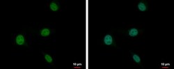
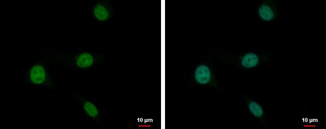
- PLZF Polyclonal Antibody detects PLZF protein at nucleus by immunofluorescent analysis. Sample: U87-MG cells were fixed in 4% paraformaldehyde at RT for 15 min. Green: PLZF protein stained by PLZF Polyclonal Antibody (Product # PA5-29213) diluted at 1:2,000. Blue: Hoechst 33342 staining.

- Submitted by
- Invitrogen Antibodies (provider)
- Main image
- Experimental details
- PLZF Polyclonal Antibody detects PLZF protein at nucleus by immunofluorescent analysis. Sample: U87-MG cells were fixed in 4% paraformaldehyde at RT for 15 min. Green: PLZF protein stained by PLZF Polyclonal Antibody (Product # PA5-29213) diluted at 1:2,000. Blue: Hoechst 33342 staining.
Supportive validation
- Submitted by
- Invitrogen Antibodies (provider)
- Main image

- Experimental details
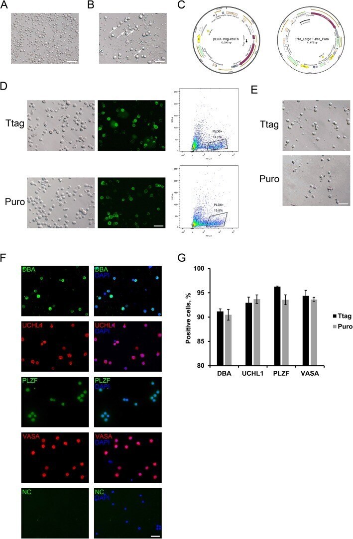
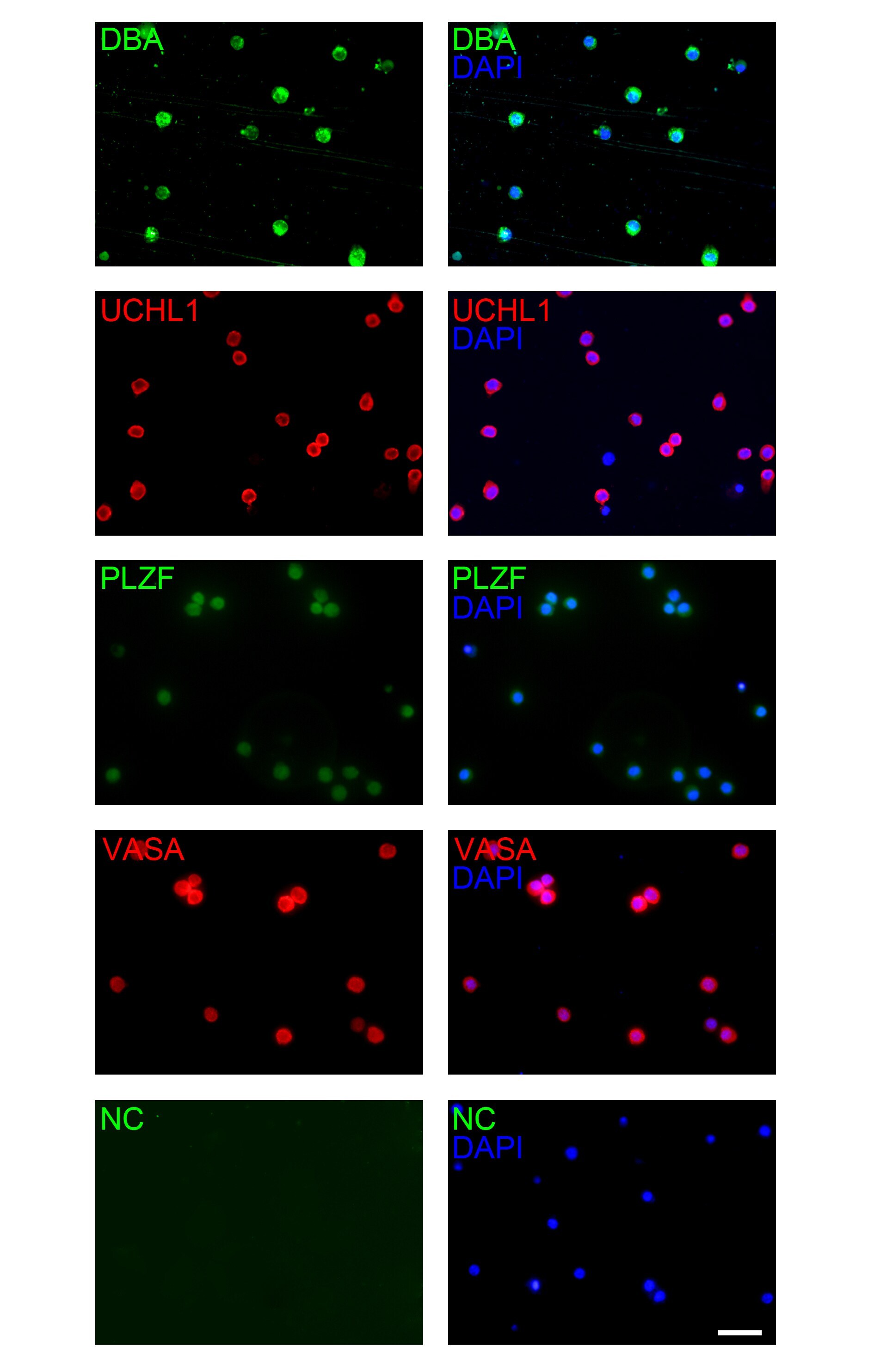
- Fig. 1 Immortalization and enrichment of porcine SSCs. a The single-cell suspension obtained after the two-enzymatic dissociation. Bar = 100 mum. b The cell fraction after differential plating. Arrows point to the putative germ cells. Bar = 100 mum. c Maps of the vector pLOX-Ttag-iresTK (hereafter referred to as Ttag) and Ef1a-Large T-Ires-Puro (hereafter referred to as Puro). d Left: living staining of Ttag- and Puro-transduced cells for PLD6. Bar = 100 mum; Right: enrichment of PLD6 + cells by FACS. e Images of Ttag- and Puro-transduced cells after sorting. Bar = 100 mum. f Staining of Ttag-transduced PLD6 + cells for lectin DBA, UCHL1, PLZF and VASA. NC: the negative control using the isotype IgG in place of the primary antibody. Bar = 50 mum. g Percentages of sorted cells positive for porcine SSC and germ cell markers. Data are presented as the mean +- SEM of three experiments (technical triplicates)
- Submitted by
- Invitrogen Antibodies (provider)
- Main image

- Experimental details
- Additional file 1: Figure S1. Staining of Puro-transduced PLD6 + cells for lectin DBA, UCHL1, PLZF and VASA. NC: the negative control using the isotype IgG in place of the primary antibody. Bar = 50 mum.
- Submitted by
- Invitrogen Antibodies (provider)
- Main image
- Experimental details
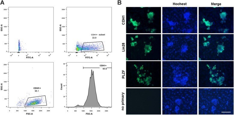
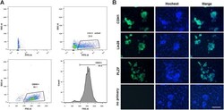
- Fig. 5 Purification and characterization of primary SSCs. a Flow cytometry data to illustrate the gating strategy for FACS purification of CDH1 + germ cells. b Immunocytochemical staining of SSC colonies on Sertoli cell feeder layer after 14 days of culture. The fluorescence staining is performed on CDH1 FACS sorted cells following differential plating. Expression of CDH1, Lin28, and PLZF in SSC colonies was showed in green fluorescence. Negative (no primary) control: omission of primary antibody. The nuclei (blue) were stained with Hoechst 33342. Scale bar: 100 mum