Explore
 Validate
 Learn
Learn Western blot
Antibody data
- Antibody Data
- Antigen structure
- References [1]
- Comments [0]
- Validations
- Western blot [1]
- Immunocytochemistry [1]
- Flow cytometry [1]
Submit
Validation data
Reference
Comment
Report error
- Product number
- MAB2944 - Provider product page

- Provider
- R&D Systems
- Product name
- Human PLZF Antibody
- Antibody type
- Monoclonal
- Description
- Protein A or G purified from hybridoma culture supernatant. Detects human PLZF in direct ELISAs and Western blots. In direct ELISAs, no cross-reactivity with recombinant human (rh) ZBTB38, rhZNF24, rhZNF143, rhZNF206, rhZNF281, or rhZNF423 is observed.
- Reactivity
- Human
- Host
- Mouse
- Conjugate
- Unconjugated
- Antigen sequence
Q05516- Isotype
- IgG
- Antibody clone number
- 6318100
- Vial size
- 100 ug
- Concentration
- LYOPH
- Storage
- Use a manual defrost freezer and avoid repeated freeze-thaw cycles. 12 months from date of receipt, -20 to -70 °C as supplied. 1 month, 2 to 8 °C under sterile conditions after reconstitution. 6 months, -20 to -70 °C under sterile conditions after reconstitution.
Submitted references PLZF, a tumor suppressor genetically lost in metastatic castration-resistant prostate cancer, is a mediator of resistance to androgen deprivation therapy.
Hsieh CL, Botta G, Gao S, Li T, Van Allen EM, Treacy DJ, Cai C, He HH, Sweeney CJ, Brown M, Balk SP, Nelson PS, Garraway LA, Kantoff PW
Cancer research 2015 May 15;75(10):1944-8
Cancer research 2015 May 15;75(10):1944-8
No comments: Submit comment
Supportive validation
- Submitted by
- R&D Systems (provider)
- Main image
- Experimental details
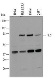
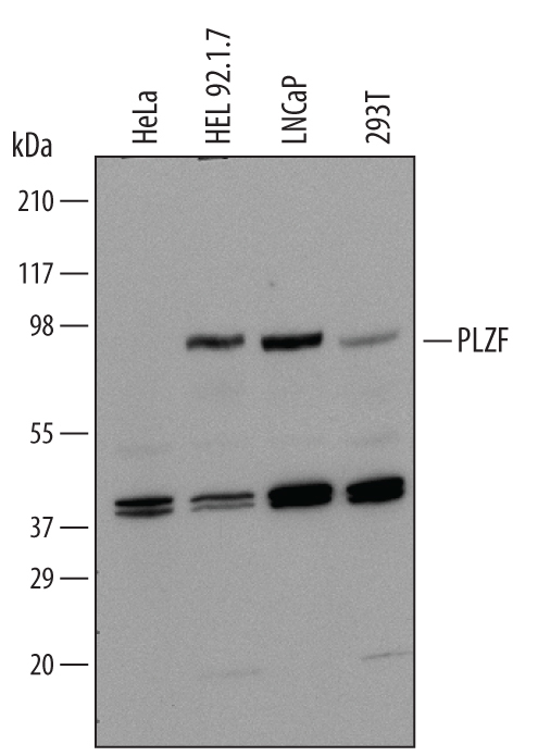
- Detection of Human PLZF by Western Blot. Western blot shows lysates of HeLa human cervical epithelial carcinoma cell line, HEL 92.1.7 human erythroleukemic cell line, LNCaP human prostate cancer cell line, and 293T human embryonic kidney cell line. PVDF Membrane was probed with 1 µg/mL of Human PLZF Monoclonal Antibody (Catalog # MAB2944) followed by HRP-conjugated Anti-Mouse IgG Secondary Antibody (Catalog # HAF007). A specific band was detected for PLZF at approximately 85 kDa (as indicated). This experiment was conducted under reducing conditions and using Immunoblot Buffer Group 1.
Supportive validation
- Submitted by
- R&D Systems (provider)
- Main image

- Experimental details
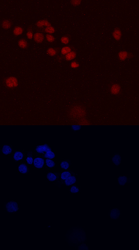
- PLZF in HL-60 Human Cell Line. PLZF was detected in immersion fixed HL-60 human acute promyelocytic leukemia cell line using Human PLZF Monoclonal Antibody (Catalog # MAB2944) at 10 µg/mL for 3 hours at room temperature. Cells were stained using the NorthernLights™ 557-conjugated Anti-Mouse IgG Secondary Antibody (red, upper panel; Catalog # NL007) and counterstained with DAPI (blue, lower panel). Specific staining was localized to nuclei. View our protocol for Fluorescent ICC Staining of Non-adherent Cells.
Supportive validation
- Submitted by
- R&D Systems (provider)
- Main image

- Experimental details
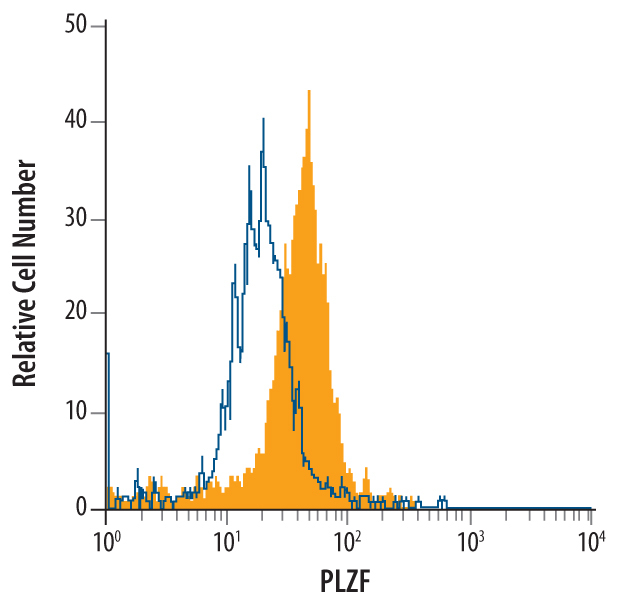
- Detection of in HL-60 Human Cell Line by Flow Cytometry. HL-60 human acute promyelocytic leukemia cell line was stained with Human PLZF Monoclonal Antibody (Catalog # MAB2944, filled histogram) or isotype control antibody (Catalog # MAB003, open histogram), followed by Phycoerythrin-conjugated Anti-Mouse IgG F(ab')2 Secondary Antibody (Catalog # F0102B). To facilitate intracellular staining, cells were fixed with paraformaldehyde and permeabilized with saponin.