 Explore
 Validate
 Learn
Learn Western blot
 Immunohistochemistry
Antibody data
- Antibody Data
- Antigen structure
- References [3]
- Comments [0]
- Validations
- Western blot [1]
- Immunocytochemistry [3]
- Immunoprecipitation [1]
Submit
Validation data
Reference
Comment
Report error
- Product number
- R1187 - Provider product page

- Provider
- Acris Antibodies GmbH
- Proper citation
- Acris Antibodies GmbH Cat#R1187, RRID:AB_1007850
- Product name
- anti TERT
- Antibody type
- Polyclonal
- Antigen
- Synthetic peptide corresponding to amino acids 1104-1123 of the carboxy terminal end of Human TERT
- Reactivity
- Human
- Host
- Rabbit
- Vial size
- 0.1 mg
- Concentration
- 1.0 mg/ml (by UV absorbance at 280 nm).
Submitted references Novel immortal human cell lines reveal subpopulations in the nucleus pulposus.
Telomerase does not counteract telomere shortening but protects mitochondrial function under oxidative stress.
Immunodetection of human telomerase reverse-transcriptase (hTERT) re-appraised: nucleolin and telomerase cross paths.
van den Akker GG, Surtel DA, Cremers A, Rodrigues-Pinto R, Richardson SM, Hoyland JA, van Rhijn LW, Welting TJ, Voncken JW
Arthritis research & therapy 2014 Jun 27;16(3):R135
Arthritis research & therapy 2014 Jun 27;16(3):R135
Telomerase does not counteract telomere shortening but protects mitochondrial function under oxidative stress.
Ahmed S, Passos JF, Birket MJ, Beckmann T, Brings S, Peters H, Birch-Machin MA, von Zglinicki T, Saretzki G
Journal of cell science 2008 Apr 1;121(Pt 7):1046-53
Journal of cell science 2008 Apr 1;121(Pt 7):1046-53
Immunodetection of human telomerase reverse-transcriptase (hTERT) re-appraised: nucleolin and telomerase cross paths.
Wu YL, Dudognon C, Nguyen E, Hillion J, Pendino F, Tarkanyi I, Aradi J, Lanotte M, Tong JH, Chen GQ, Ségal-Bendirdjian E
Journal of cell science 2006 Jul 1;119(Pt 13):2797-806
Journal of cell science 2006 Jul 1;119(Pt 13):2797-806
No comments: Submit comment
Supportive validation
- Submitted by
- Acris Antibodies GmbH (provider)
- Main image

- Experimental details
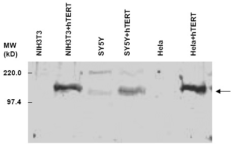
- Figure 1. Immunoblot using anti-hTERT antibody (1/500 dilution). Endogenous levels of mTERT in NIH 3T3 cells (lane 1) and hTERT in HeLa cells (lane 5) are not detectable. After transduction with an hTERT expression vector, both cell types show high levels of hTERT protein (lanes 2 and 6). SY5Y cells, which have high endogenous levels of hTERT, have detectable hTERT protein in both untransduced (lane 3) and transduced (lane 4) cells. The arrow indicates a molecular weight of approximately 127kD, the expected size of hTERT protein.
Supportive validation
- Submitted by
- Acris Antibodies GmbH (provider)
- Main image
- Experimental details
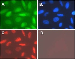
- Figure 3. Immunofluorescence microscopy of Saos-2 cells transduced with a retroviral vector expressing hTERT and green fluorescent protein (GFP) from an internal ribosomal entry site (IRES). Panel A shows native GFP expression (green), Panel B shows DAPI staining of chromsomes (blue), Panel C shows anti hTERT staining at a 1/500 dilution followed by washes and addition of a 1/1000 dilution of Rhodamine Goat anti-Rabbit IgG (Cat.-No R1364T) for detection. Panel D shows no staining of hTERT-transduced Cells using pre-immune serum.

- Submitted by
- Acris Antibodies GmbH (provider)
- Main image
- Experimental details
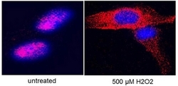
- Figure 4. Immunofluorescence using Anti Human TERT Antibody Cat.-No R1187 to stain hTERT on hTERT-over-expressing fibroblasts. Cells were untreated (Left) or treated (Right) with 500 uM H202, fixed in 4% PFA (in PBS) for 10 min and frozen in -80 after 3 min air-drying before staining with  Anti Human TERT Antibody Cat.-No R1187 at 1/2000 overnight. Confocal images provided by G. Saretzki, Institute for Ageing and Health, Newcastle University, UK. See Ahmed et. Al. for more information.

- Submitted by
- Acris Antibodies GmbH (provider)
- Main image

- Experimental details
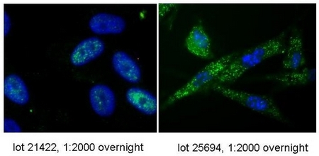
- Figure 5. Immunofluorescence using Two different Lots of Anti Human TERT Antibody Cat.-No R1187 to stain hTERT on hTERT-over-expressing fibroblasts. Cells were fixed in 4% PFA (in PBS) for 10 min and frozen in -80 after 3 min air-drying before incubation with Anti Human TERT Antibody Cat.-No R1187 at 1/2000 overnight and staining with a 1/2000 dilution of Alexafluor488 secondary Ab. Confocal images provided by G. Saretzki, Institute for Ageing and Health, Newcastle University, UK. See Ahmed et. Al. for more information.
Supportive validation
- Submitted by
- Acris Antibodies GmbH (provider)
- Main image
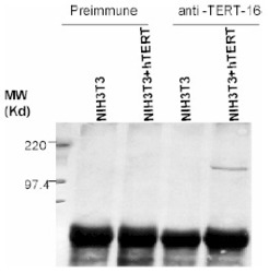
- Experimental details
- Figure 2. Immunoprecipitation of hTERT protein from NIH 3T3 cell lysates. The anti-hTERT antibody was used for both immunoprecipitation and immunoblotting. Anti-hTERT antibody was able to immunoprecipitate TERT protein from cells with ectopic hTERT expression (lane 4). The preimmune serum was unable to immunoprecipitate TERT protein (Lanes 1 and 2).
