Explore
 Validate
 Learn
Learn Western blot
 ELISA
ELISAAntibody data
- Antibody Data
- Antigen structure
- References [0]
- Comments [0]
- Validations
- Western blot [2]
- Immunohistochemistry [9]
Submit
Validation data
Reference
Comment
Report error
- Product number
- ABIN2506515 - Provider product page

- Provider
- antibodies-online
- Product name
- anti-Telomerase Reverse Transcriptase (TERT) (Ser842) antibody
- Antibody type
- Polyclonal
- Antigen
- The antiserum was produced against synthesized non-phosphopeptide derived from human TERT around the Ser842 (G-K-SP-Y-V).
- Description
- The antibody was affinity-purified from rabbit antiserum by affinity-chromatography using epitope-specific immunogen.
- Reactivity
- Human
- Host
- Rabbit
- Epitope
- Ser842
- Isotype
- IgG
- Vial size
- 100 μg
- Storage
- Store at -20°C/1 year
No comments: Submit comment
Supportive validation
- Submitted by
- antibodies-online (provider)
- Main image
- Experimental details
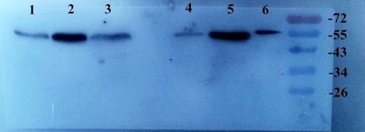
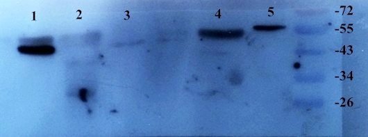
- WB analysis of Rat lung (lane 1), Rat heart (lane 2), Rat liver (lane 3), Mouse spinal cord (lane 4), Rat kidney (lane 5), Mouse large intestine (lane 6) using TGF beta 1 antibody primary antibody at (2 ug/ml)
- Submitted by
- antibodies-online (provider)
- Main image
- Experimental details
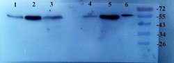
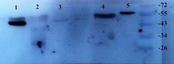
- Western blot analysis of Rat heart (lane 1), Rat liver (lane 2), Rat brain (lane 3), Rat kidney (lane 4), Mouse large intestine (lane 5) TGF beta 1 antibody dilution of primary antibody - (2 ug/ml)
Supportive validation
- Submitted by
- antibodies-online (provider)
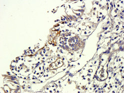
- Main image
- Experimental details
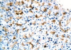
- Rat kidney cells expressing TGF beta 1 (IHC-p with DAB as chromogen)
- Submitted by
- antibodies-online (provider)
- Main image
- Experimental details
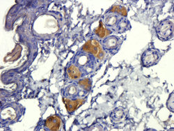
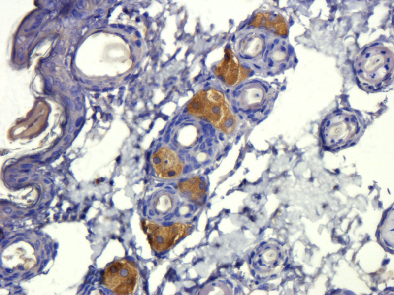
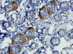
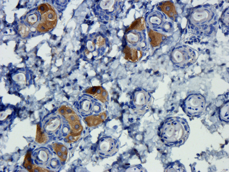
- Immunohistochemical staining of mouse skin tissue using TGF beta 1 antibody (dilution of primary antibody - 1:300)

- Submitted by
- antibodies-online (provider)
- Main image
- Experimental details
- Immunohistochemical staining of pig large intestines tissue using anti-TGF beta 1 (dilution of primary antibody - 1:300)

- Submitted by
- antibodies-online (provider)
- Main image
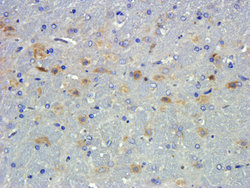
- Experimental details
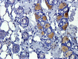
- IHC-P image of rat brain tissue using anti-TGF beta 1 (dilution of primary antibody at 1:300)

- Submitted by
- antibodies-online (provider)
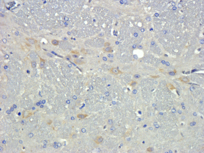
- Main image

- Experimental details
- IHC-P image of rat brain tissue using TGF beta 1 antibody (dilution of primary antibody at 1:300)
- Submitted by
- antibodies-online (provider)
- Main image
- Experimental details
- Immunohistochemical staining of mouse skin tissue using TGF beta 1 antibody (dilution of primary antibody - 1:800)

- Submitted by
- antibodies-online (provider)
- Main image
- Experimental details
- Immunohistochemical staining of paraffin embedded mouse skin tissue using anti-TGF beta 1 (primary antibody at 1:300)
- Submitted by
- antibodies-online (provider)
- Main image
- Experimental details
- IHC-P image of mouse skin tissue using TGF beta 1 antibody (dilution of primary antibody at 1:600)
- Submitted by
- antibodies-online (provider)
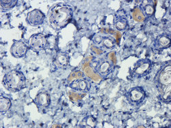
- Main image
- Experimental details
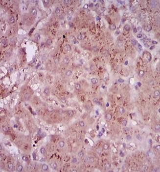
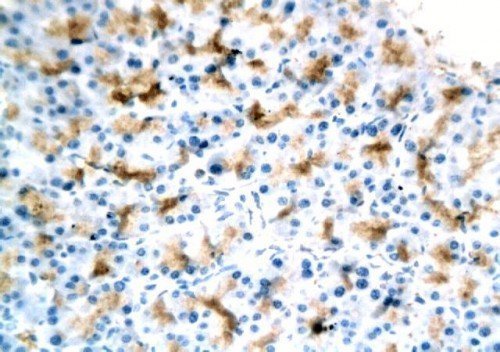
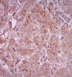
- Immunohistochemical analysis of paraffin-embedded human liver tissue using tgf-beta antibody(5ug/ml)