Explore
 Validate
 Learn
Learn Western blot
Antibody data
- Antibody Data
- Antigen structure
- References [0]
- Comments [0]
- Validations
- Western blot [1]
- Immunocytochemistry [2]
- Immunohistochemistry [1]
- Flow cytometry [1]
Submit
Validation data
Reference
Comment
Report error
- Product number
- AP33476PU-N - Provider product page

- Provider
- OriGene
- Product name
- Telomerase reverse transcriptase (TERT) (627-656) rabbit polyclonal antibody, Purified
- Antibody type
- Polyclonal
- Description
- Telomerase reverse transcriptase (TERT) (627-656) rabbit polyclonal antibody, Purified
- Host
- Rabbit
- Conjugate
- Unconjugated
- Epitope
- TERT
- Antibody clone number
- NULL
- Vial size
- 200 µl
- Concentration
- 2.0 mg/ml (lot specific)
No comments: Submit comment
Supportive validation
- Submitted by
- OriGene (provider)
- Main image

- Experimental details
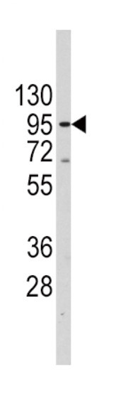
- Western blot of anti-TERT Antibody in Jurkat cell line lysates (35 ug/lane). TERT(arrow) was detected using the purified antibody.
- Validation comment
- WB
Supportive validation
- Submitted by
- OriGene (provider)
- Main image
- Experimental details
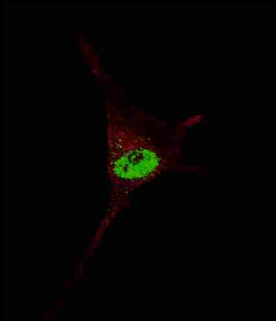
- Fluorescent confocal image of SY5Y cells stained with TERT antibody. SY5Y cells were fixed with 4% PFA (20 min), permeabilized with Triton X-100 (0.2%, 30 min). Cells were then incubated TERT primary antibody (1:200, 2 h at room temperature). For secondary antibody, Alexa Fluor 488 conjugated donkey anti-rabbit antibody (green) was used (1:1000, 1h). Nuclei were counterstained with Hoechst 33342 (blue) (10 ug/ml, 5 min). TERT immunoreactivity is localized to the nucleus of SY5Y cells.
- Validation comment
- IF
- Submitted by
- OriGene (provider)
- Main image

- Experimental details
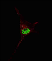
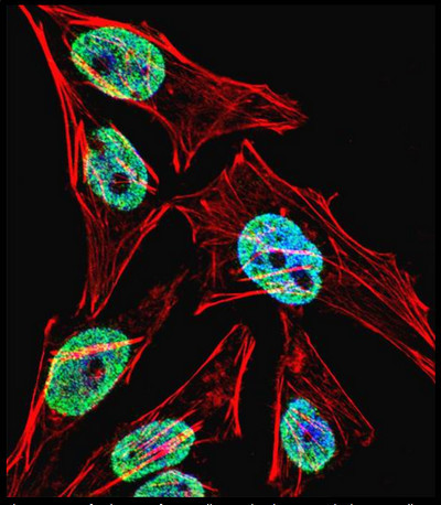
- Fluorescent confocal image of HeLa cell stained with TERT Antibody. HeLa cells were fixed with 4% PFA (20 min), permeabilized with Triton X-100 (0.1%, 10 min), then incubated with TERT primary antibody (1:25, 1 h at 37°C). For secondary antibody, Alexa Fluor 488 conjugated donkey anti-rabbit antibody (green) was used (1:400, 50 min at 37°C). Cytoplasmic actin was counterstained with Alexa Fluor 555 (red) conjugated Phalloidin (7units/ml, 1 h at 37°C). Nuclei were counterstained with DAPI (blue) (10 ug/ml, 10 min). TERT immunoreactivity is localized to Nucleus significantly and Cytoplasm weakly.
- Validation comment
- IF
Supportive validation
- Submitted by
- OriGene (provider)
- Main image

- Experimental details
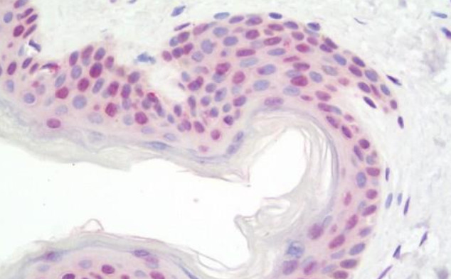
- Formalin-Fixed, Paraffin-Embedded Human Skin tissue stained with Anti-TERT / Telomerase antibody ? at 1/100 dilution after heat-induced antigen retrieval.
- Validation comment
- IHC
Supportive validation
- Submitted by
- OriGene (provider)
- Main image
- Experimental details
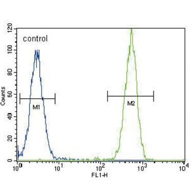
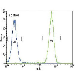
- TERT Antibody flow cytometry of HeLa cells (right histogram) compared to a negative control cell (left histogram). FITC-conjugated goat-anti-rabbit secondary antibodies were used for the analysis.
- Validation comment
- FC