 Explore
 Validate
 Learn
Learn Western blot
 Immunocytochemistry
Antibody data
- Antibody Data
- Antigen structure
- References [1]
- Comments [0]
- Validations
- Immunocytochemistry [7]
- Immunohistochemistry [1]
- Flow cytometry [1]
Submit
Validation data
Reference
Comment
Report error
- Product number
- MA5-16033 - Provider product page

- Provider
- Invitrogen Antibodies
- Product name
- TERT Monoclonal Antibody (2D8)
- Antibody type
- Monoclonal
- Antigen
- Recombinant full-length protein
- Description
- Suggested positive control: 293T whole cell lysates, HeLa whole cell lysates, antigen standard for TERT (transient overexpression lysate).
- Reactivity
- Human
- Host
- Mouse
- Isotype
- IgM
- Antibody clone number
- 2D8
- Vial size
- 100 μL
- Concentration
- Conc. Not Determined
- Storage
- -20°C or -80°C if preferred
Submitted references Clinicopathological Analysis of HIF-1alpha and TERT on Survival Outcome in Glioblastoma Patients: A Prospective, Single Institution Study.
Potharaju M, Mathavan A, Mangaleswaran B, Patil S, John R, Ghosh S, Kalavakonda C, Ghosh M, Verma RS
Journal of Cancer 2019;10(11):2397-2406
Journal of Cancer 2019;10(11):2397-2406
No comments: Submit comment
Supportive validation
- Submitted by
- Invitrogen Antibodies (provider)
- Main image

- Experimental details
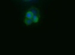
- Immunocytochemistry analysis of TERT in HepG2 cells. Samples were incubated in TERT monoclonal antibody (Product # MA5-16033). Tert (green). Nuclei (Blue) are counterstained with Hoechst 33258.

- Submitted by
- Invitrogen Antibodies (provider)
- Main image

- Experimental details
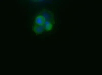
- Immunocytochemistry analysis of TERT in HepG2 cells. Samples were incubated in TERT monoclonal antibody (Product # MA5-16033). Tert (green). Nuclei (Blue) are counterstained with Hoechst 33258.

- Submitted by
- Invitrogen Antibodies (provider)
- Main image
- Experimental details
- Immunocytochemistry analysis of TERT in HepG2 cells. Samples were incubated in TERT monoclonal antibody (Product # MA5-16033). Tert (green). Nuclei (Blue) are counterstained with Hoechst 33258.
- Submitted by
- Invitrogen Antibodies (provider)
- Main image

- Experimental details
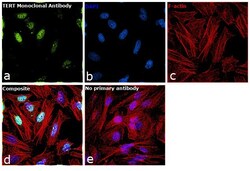
- Immunofluorescence analysis of TERT was performed using 70% confluent log phase HeLa cells. The cells were fixed with 4% paraformaldehyde for 10 minutes, permeabilized with 0.1% Triton™ X-100 for 15 minutes, and blocked with 1% BSA for 1 hour at room temperature. The cells were labeled with Anti-TERT Mouse Monoclonal Antibody (2D8) (Product # MA5-16033) at 1:200 dilution in 0.1% BSA, incubated at 4 degree Celsius overnight and then labeled with Goat anti-Mouse IgG (H+L)/IgM (L) Superclonal™ Secondary Antibody, Alexa Fluor® 488 conjugate (Product # A28175) at a dilution of 1:2000 for 45 minutes at room temperature (Panel a: green).Nuclei (Panel b: blue) were stained with ProLong™ Diamond Antifade Mountant with DAPI (Product # P36962). F-actin (Panel c: red) was stained with Rhodamine Phalloidin (Product # R415, 1:300). Panel d represents the merged image showing nuclear localization. Panel e shows cells with no primary antibody to assess background. The images were captured at 60X magnification.

- Submitted by
- Invitrogen Antibodies (provider)
- Main image
- Experimental details
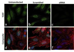
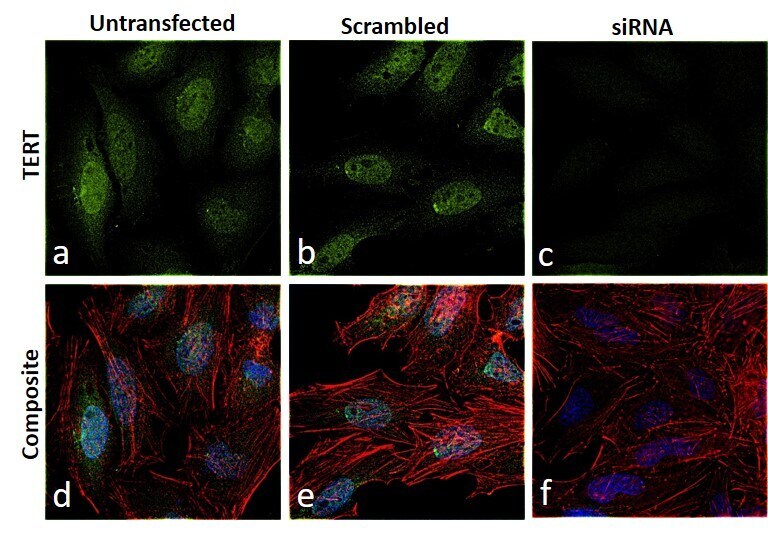
- Knockdown of TERT was achieved by transfecting HeLa cells with TERT specific siRNA (Silencer® select Product # s370, s371). Immunofluorescence analysis was performed on HeLa cells (untransfected, panel a,d), transfected with non-specific scrambled siRNA (panels b,e) and transfected with TERT specific siRNA (panel c,f) Cells were fixed, permeabilized, and labelled with TERT Monoclonal Antibody (2D8) (Product # MA5-16033, 1:200 dilution), followed by Goat anti-Mouse IgG (H+L)/IgM (L) Superclonal™ Secondary Antibody, Alexa Fluor® 488 conjugate (Product # A28175, 1:2000). Nuclei (blue) were stained using ProLong™ Diamond Antifade Mountant with DAPI (Product # P36962), and Rhodamine Phalloidin (Product # R415, 1:300) was used for cytoskeletal F-actin (red) staining. Reduction of specific signal was observed upon siRNA mediated knockdown (panel c,f) confirming specificity of the antibody to TERT (green). The images were captured at 60X magnification.
- Submitted by
- Invitrogen Antibodies (provider)
- Main image

- Experimental details
- Knockdown of TERT was achieved by transfecting HeLa cells with TERT specific siRNA (Silencer® select Product # s370, s371). Immunofluorescence analysis was performed on HeLa cells (untransfected, panel a,d), transfected with non-specific scrambled siRNA (panels b,e) and transfected with TERT specific siRNA (panel c,f) Cells were fixed, permeabilized, and labelled with TERT Monoclonal Antibody (2D8) (Product # MA5-16033, 1:200 dilution), followed by Goat anti-Mouse IgG (H+L)/IgM (L) Superclonal™ Secondary Antibody, Alexa Fluor® 488 conjugate (Product # A28175, 1:2000). Nuclei (blue) were stained using ProLong™ Diamond Antifade Mountant with DAPI (Product # P36962), and Rhodamine Phalloidin (Product # R415, 1:300) was used for cytoskeletal F-actin (red) staining. Reduction of specific signal was observed upon siRNA mediated knockdown (panel c,f) confirming specificity of the antibody to TERT (green). The images were captured at 60X magnification.

- Submitted by
- Invitrogen Antibodies (provider)
- Main image
- Experimental details
- Immunofluorescence analysis of TERT was performed using 70% confluent log phase HeLa cells. The cells were fixed with 4% paraformaldehyde for 10 minutes, permeabilized with 0.1% Triton™ X-100 for 15 minutes, and blocked with 1% BSA for 1 hour at room temperature. The cells were labeled with Anti-TERT Mouse Monoclonal Antibody (2D8) (Product # MA5-16033) at 1:200 dilution in 0.1% BSA, incubated at 4 degree Celsius overnight and then labeled with Goat anti-Mouse IgG (H+L)/IgM (L) Superclonal™ Secondary Antibody, Alexa Fluor® 488 conjugate (Product # A28175) at a dilution of 1:2000 for 45 minutes at room temperature (Panel a: green).Nuclei (Panel b: blue) were stained with ProLong™ Diamond Antifade Mountant with DAPI (Product # P36962). F-actin (Panel c: red) was stained with Rhodamine Phalloidin (Product # R415, 1:300). Panel d represents the merged image showing nuclear localization. Panel e shows cells with no primary antibody to assess background. The images were captured at 60X magnification.
Supportive validation
- Submitted by
- Invitrogen Antibodies (provider)
- Main image
- Experimental details
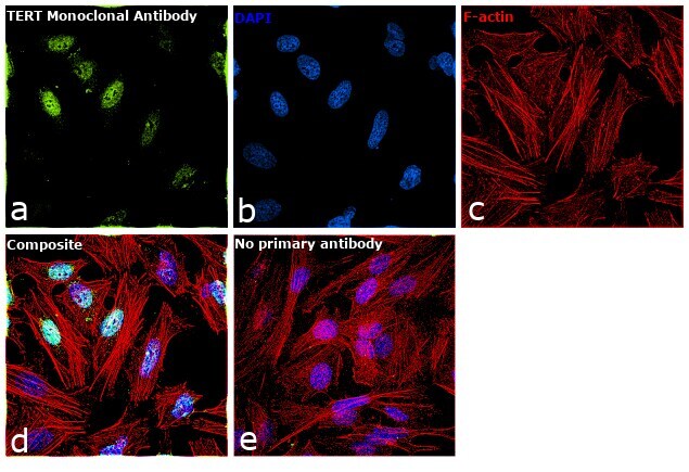
- Immunohistochemical analysis of TERT in immersion fixed paraffin-embedded sections of human liver cancer. Samples were incubated in TERT monoclonal antibody (Product # MA5-16033) using a dilution of 1:600 overnight at 4 °C. Tissue was stained using the VisuCyte anti-mouse HRP polymer detection reagent with DAB chromogen (brown) and counterstained with hematoxylin (blue).

Supportive validation
- Submitted by
- Invitrogen Antibodies (provider)
- Main image
- Experimental details
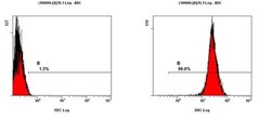
- Flow cytometry analysis of TERT using a monoclonal antibody (Product # MA5-16033).
