Explore
 Validate
 Learn
Learn Western blot
Antibody data
- Antibody Data
- Antigen structure
- References [1]
- Comments [0]
- Validations
- Western blot [2]
- Immunocytochemistry [1]
- Chromatin Immunoprecipitation [1]
- Other assay [2]
Submit
Validation data
Reference
Comment
Report error
- Product number
- 712089 - Provider product page

- Provider
- Invitrogen Antibodies
- Product name
- YY1 Recombinant Polyclonal Antibody (6HCLC)
- Antibody type
- Polyclonal
- Antigen
- Other
- Reactivity
- Human
- Host
- Rabbit
- Isotype
- IgG
- Antibody clone number
- 6HCLC
- Vial size
- 100 µg
- Concentration
- 0.5 mg/mL
- Storage
- Store at 4°C short term. For long term storage, store at -20°C, avoiding freeze/thaw cycles.
Submitted references Long noncoding RNA LINC01578 drives colon cancer metastasis through a positive feedback loop with the NF-κB/YY1 axis.
Liu J, Zhan Y, Wang J, Wang J, Guo J, Kong D
Molecular oncology 2020 Dec;14(12):3211-3233
Molecular oncology 2020 Dec;14(12):3211-3233
No comments: Submit comment
Supportive validation
- Submitted by
- Invitrogen Antibodies (provider)
- Main image
- Experimental details
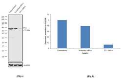
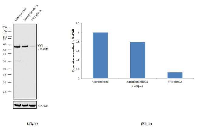
- Knockdown of YY1 was achieved by transfecting DU 145 cells with YY1 specific siRNA (Silencer® select Product # s14958 & s14960). Western blot analysis (Fig a) was performed using modified whole cell extracts (1% SDS) from YY1 knockdown cells (Lane 3), non-specific scrambled siRNA transfected cells (Lane 2) and untransfected cells (Lane 1). The blot was probed with Anti-YY1 Recombinant Rabbit Polyclonal Antibody (Product # 712089, 1:500 dilution) and Goat anti-Rabbit IgG (H+L) Superclonal™ Secondary Antibody, HRP conjugate (Product # A27036, 0.25 µg/mL, 1:4000 dilution). Densitometric analysis of this Western blot is shown in histogram (Fig b). Loss of signal upon siRNA mediated knockdown confirms that antibody is specific to YY1.
- Submitted by
- Invitrogen Antibodies (provider)
- Main image
- Experimental details
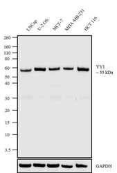
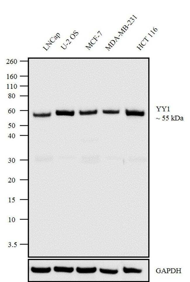
- Western blot analysis was performed on modified nuclear extracts (1% SDS) (30 µg lysate) of LNCap (Lane 1), U-2 OS (Lane 2), MCF-7 (Lane 3), MDA-MB-231 (Lane 4), and HCT 116 (Lane 5). The blot was probed with Anti-YY1 Recombinant Rabbit Polyclonal Antibody (Product # 712089, 1:500 dilution) and detected by chemiluminescence using Goat anti-Rabbit IgG (H+L) Superclonal™ Secondary Antibody, HRP conjugate (Product # A27036, 0.25 µg/ml, 1:4000 dilution). A 55 kDa band corresponding to YY1 was observed across the cell lines tested.
Supportive validation
- Submitted by
- Invitrogen Antibodies (provider)
- Main image
- Experimental details
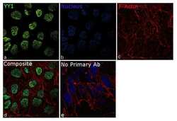
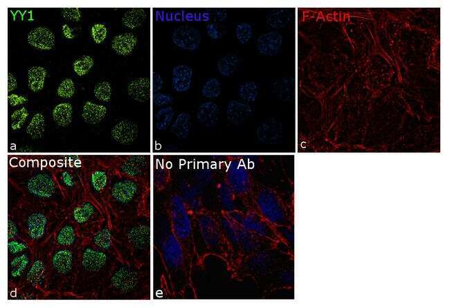
- For immunofluorescence analysis, DU 145 cells were fixed and permeabilized for detection of endogenous YY1 using Anti-YY1 Recombinant Rabbit Polyclonal Antibody (Product # 712089, 1:100 dilution) and labeled with Goat anti-Rabbit IgG (H+L) Superclonal™ Secondary Antibody, Alexa Fluor® 488 conjugate (Product # A27034, 1:2000). Panel a) shows representative cells that were stained for detection and localization of YY1 protein (green), Panel b) is stained for nuclei (blue) using ProLong™ Diamond Antifade Mountant with DAPI (Product # P36962). Panel c) represents cytoskeletal F-actin staining using Rhodamine Phalloidin (Product # R415, 1:300). Panel d) is a composite image of Panels a, b and c clearly demonstrating nuclear localization of YY1. Panel e) represents control cells with no primary antibody to assess background. The images were captured at 60X magnification.
Supportive validation
- Submitted by
- Invitrogen Antibodies (provider)
- Main image
- Experimental details
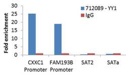
- Enrichment of endogenous YY1 protein at specific gene loci using Anti-YY1 Antibody: Chromatin Immunoprecipitation (ChIP) was performed using Anti-YY1 Recombinant Rabbit Polyclonal Antibody (Product # 712089, 4 µg) on sheared chromatin from 2 million U-2 OS cells using the MAGnify ChIP System kit (Product # 49-2024). Normal Rabbit IgG was used as a negative IP control. The purified DNA was analyzed by qPCR with PCR primer pairs over the promoters of CXXC1, FAM193B (active) and SAT2 and SAT alpha satellite repeats (inactive). Data is presented as fold enrichment of the antibody signal versus the negative control IgG using the comparative CT method.

Supportive validation
- Submitted by
- Invitrogen Antibodies (provider)
- Main image
- Experimental details
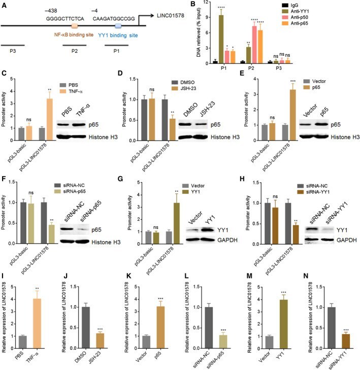
- Fig. 2 LINC01578 is upregulated by NF-kappaB and YY1. (A) The predicted NF-kappaB and YY1 binding sites on LINC01578 promoter. NF-kappaB and YY1 bind sites were at -438 and -4 positions relative to the transcription start site, respectively. (B) ChIP assays were performed in DLD-1 cells to measure the binding of NF-kappaB and YY1 on LINC01578 promoter. A distant region without NF-kappaB and YY1 binding sites was used as NC (P3). (C) Luciferase reporter assays for DLD-1 cells transfected with luciferase reporter plasmids containing LINC01578 promoter and treated with PBS or 10 ng*mL -1 TNF-alpha for 24 h. Nuclear p65 levels of DLD-1 cell treatment with PBS or 10 ng*mL -1 TNF-alpha for 24 h were detected by western blot. (D) Luciferase reporter assays for DLD-1 cells transfected with luciferase reporter plasmids containing LINC01578 promoter and treated with DMSO or 5 u m JSH-23 for 24 h. Nuclear p65 levels of DLD-1 cell treatment with DMSO or 5 u m JSH-23 for 24 h were detected by western blot. (E) Luciferase reporter assays for DLD-1 cells cotransfected with luciferase reporter plasmids containing LINC01578 promoter and p65 overexpression vector. p65 overexpression efficiencies were detected by western blot. (F) Luciferase reporter assays for DLD-1 cells cotransfected with luciferase reporter plasmids containing LINC01578 promoter and siRNAs against p65. p65 knockdown efficiencies were detected by western blot. (G) Luciferase reporter assays for DLD-1 cells cotransfected with
- Submitted by
- Invitrogen Antibodies (provider)
- Main image

- Experimental details
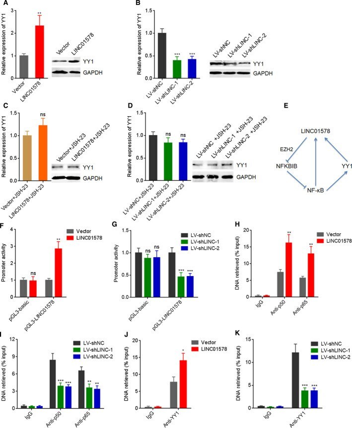
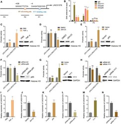
- Fig. 9 LINC01578 forms a positive feedback loop with NF-kappaB/YY1. (A) YY1 mRNA and protein levels in DLD-1 cells transfected with LINC01578 overexpression vector were determined by qRT-PCR and western blot. (B) YY1 mRNA and protein levels in LoVo cells infected with shRNAs targeted to LINC01578 were determined by qRT-PCR and western blot. (C) YY1 mRNA and protein levels in DLD-1 cells transfected with LINC01578 overexpression vector and treated with 5 u m JSH-23 were determined by qRT-PCR and western blot. (D) YY1 mRNA and protein levels in LoVo cells infected with shRNAs targeted to LINC01578 and treated with 5 u m JSH-23 were determined by qRT-PCR and western blot. (E) A schematic model of positive feedback loop between LINC01578 and NF-kappaB/YY1. (F) Luciferase reporter assays for LINC01578-overexpressed and control DLD-1 cells transfected with luciferase reporter plasmids containing LINC01578 promoter. (G) Luciferase reporter assays for LINC01578-depleted and control LoVo cells transfected with luciferase reporter plasmids containing LINC01578 promoter. (H) ChIP assays using p50 and p65 antibodies were carried out in LINC01578-overexpressed and control DLD-1 cells. The enrichment of LINC01578 promoter was determined by qPCR. (I) ChIP assays using p50 and p65 antibodies were carried out in LINC01578-depleted and control LoVo cells. The enrichment of LINC01578 promoter was determined by qPCR. (J) ChIP assays using YY1 antibody were carried out in LINC01578-overexpressed