Explore
 Validate
 Learn
Learn Western blot
Antibody data
- Antibody Data
- Antigen structure
- References [0]
- Comments [0]
- Validations
- Western blot [1]
- Immunohistochemistry [1]
Submit
Validation data
Reference
Comment
Report error
- Product number
- AF3784 - Provider product page

- Provider
- R&D Systems
- Product name
- Human YY1 Antibody
- Antibody type
- Polyclonal
- Description
- Immunogen affinity purified. Detects human YY1 in Western blots.
- Reactivity
- Human
- Host
- Goat
- Conjugate
- Unconjugated
- Antigen sequence
P25490- Isotype
- IgG
- Vial size
- 100 ug
- Concentration
- LYOPH
- Storage
- Use a manual defrost freezer and avoid repeated freeze-thaw cycles. 12 months from date of receipt, -20 to -70 °C as supplied. 1 month, 2 to 8 °C under sterile conditions after reconstitution. 6 months, -20 to -70 °C under sterile conditions after reconstitution.
No comments: Submit comment
Supportive validation
- Submitted by
- R&D Systems (provider)
- Main image
- Experimental details
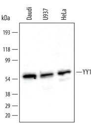
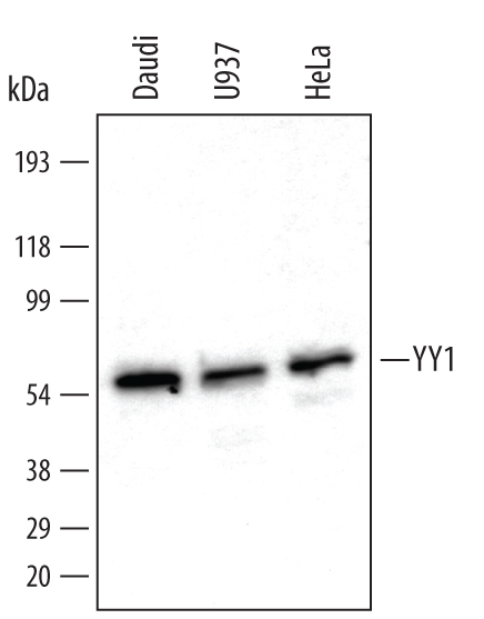
- Detection of Human YY1 by Western Blot. Western blot shows nuclear extracts of Daudi human Burkitt's lymphoma cell line, U937 human histiocytic lymphoma cell line, and HeLa human cervical epithelial carcinoma cell line. PVDF membrane was probed with 0.5 µg/mL of Goat Anti-Human YY1 Antigen Affinity-purified Polyclonal Antibody (Catalog # AF3784) followed by HRP-conjugated Anti-Goat IgG Secondary Antibody (Catalog # HAF109). A specific band was detected for YY1 at approximately 56 kDa (as indicated). This experiment was conducted under reducing conditions and using Immunoblot Buffer Group 1.
Supportive validation
- Submitted by
- R&D Systems (provider)
- Main image
- Experimental details
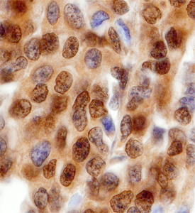
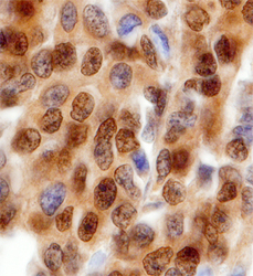
- YY1 in Human Breast. YY1 was detected in immersion fixed paraffin-embedded sections of human breast using Goat Anti-Human YY1 Antigen Affinity-purified Polyclonal Antibody (Catalog # AF3784) at 1 µg/mL overnight at 4 °C. Before incubation with the primary antibody, tissue was subjected to heat-induced epitope retrieval using Antigen Retrieval Reagent-Basic (Catalog # CTS013). Tissue was stained using the Anti-Goat HRP-DAB Cell & Tissue Staining Kit (brown; Catalog # CTS008) and counter-stained with hematoxylin (blue). Specific staining was localized to nuclei. View our protocol for Chromogenic IHC Staining of Paraffin-embedded Tissue Sections.