Explore
 Validate
 Learn
Learn Western blot
Antibody data
- Antibody Data
- Antigen structure
- References [2]
- Comments [0]
- Validations
- Western blot [2]
- Immunocytochemistry [1]
- Immunohistochemistry [1]
- Flow cytometry [1]
Submit
Validation data
Reference
Comment
Report error
- Product number
- MAB45341 - Provider product page

- Provider
- R&D Systems
- Product name
- Human FoxP1 Antibody
- Antibody type
- Monoclonal
- Description
- Protein A or G purified from hybridoma culture supernatant. Detects human FoxP1 in direct ELISAs and Western blots.
- Reactivity
- Human
- Host
- Mouse
- Conjugate
- Unconjugated
- Antigen sequence
Q9H334- Isotype
- IgG
- Antibody clone number
- 837016
- Vial size
- 100 ug
- Concentration
- LYOPH
- Storage
- Use a manual defrost freezer and avoid repeated freeze-thaw cycles. 12 months from date of receipt, -20 to -70 °C as supplied. 1 month, 2 to 8 °C under sterile conditions after reconstitution. 6 months, -20 to -70 °C under sterile conditions after reconstitution.
Submitted references Cell cycle inhibitors protect motor neurons in an organoid model of Spinal Muscular Atrophy.
Regulated expression of the TPβ isoform of the human T prostanoid receptor by the tumour suppressors FOXP1 and NKX3.1: Implications for the role of thromboxane in prostate cancer.
Hor JH, Soh ES, Tan LY, Lim VJW, Santosa MM, Winanto, Ho BX, Fan Y, Soh BS, Ng SY
Cell death & disease 2018 Oct 27;9(11):1100
Cell death & disease 2018 Oct 27;9(11):1100
Regulated expression of the TPβ isoform of the human T prostanoid receptor by the tumour suppressors FOXP1 and NKX3.1: Implications for the role of thromboxane in prostate cancer.
O'Sullivan AG, Eivers SB, Mulvaney EP, Kinsella BT
Biochimica et biophysica acta. Molecular basis of disease 2017 Dec;1863(12):3153-3169
Biochimica et biophysica acta. Molecular basis of disease 2017 Dec;1863(12):3153-3169
No comments: Submit comment
Supportive validation
- Submitted by
- R&D Systems (provider)
- Main image
- Experimental details
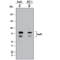
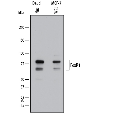
- Detection of Human FoxP1 by Western Blot. Western blot shows lysates of Daudi human Burkitt's lymphoma cell line and MCF-7 human breast cancer cell line. Gels were loaded with 25 µg of whole cell lysate (WCL) and 25 µg of nuclear extracts (Nuc). PVDF membrane was probed with 1 µg/mL of Mouse Anti-Human FoxP1 Monoclonal Antibody (Catalog # MAB45341) followed by HRP-conjugated Anti-Mouse IgG Secondary Antibody (Catalog # HAF018). Specific bands were detected for FoxP1 at approximately 65 and 80 kDa (as indicated). This experiment was conducted under reducing conditions and using Immunoblot Buffer Group 1.
- Submitted by
- R&D Systems (provider)
- Main image
- Experimental details
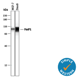
- Detection of Human FoxP1 by Simple WesternTM. Simple Western lane view shows lysates of MCF-7 human breast cancer cell line and Daudi human Burkitt's lymphoma cell line, loaded at 0.5 mg/mL. A specific band was detected for FoxP1 at approximately 99 kDa (as indicated) using 10 µg/mL of Mouse Anti-Human FoxP1 Monoclonal Antibody (Catalog # MAB45341). This experiment was conducted under reducing conditions and using the 12-230 kDa separation system.

Supportive validation
- Submitted by
- R&D Systems (provider)
- Main image
- Experimental details
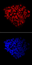
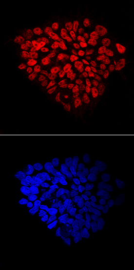
- FoxP1 in BG01V Human Embryonic Stem Cells. FoxP1 was detected in immersion fixed BG01V human embryonic stem cells using Mouse Anti-Human FoxP1 Monoclonal Antibody (Catalog # MAB45341) at 10 µg/mL for 3 hours at room temperature. Cells were stained using the NorthernLights™ 557-conjugated Anti-Mouse IgG Secondary Antibody (red, upper panel; Catalog # NL007) and counterstained with DAPI (blue, lower panel). Specific staining was localized to nuclei. View our protocol for Fluorescent ICC Staining of Stem Cells on Coverslips.
Supportive validation
- Submitted by
- R&D Systems (provider)
- Main image
- Experimental details
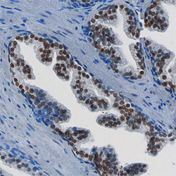
- FoxP1 in Human Prostate. FoxP1 was detected in immersion fixed paraffin-embedded sections of human prostate using Mouse Anti-Human FoxP1 Monoclonal Antibody (Catalog # MAB45341) at 15 µg/mL overnight at 4 °C. Before incubation with the primary antibody, tissue was subjected to heat-induced epitope retrieval using Antigen Retrieval Reagent-Basic (Catalog # CTS013). Tissue was stained using the Anti-Mouse HRP-DAB Cell & Tissue Staining Kit (brown; Catalog # CTS002) and counter-stained with hematoxylin (blue). Specific staining was localized to the nuclei of epithelial cells. View our protocol for Chromogenic IHC Staining of Paraffin-embedded Tissue Sections.

Supportive validation
- Submitted by
- R&D Systems (provider)
- Main image
- Experimental details
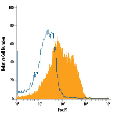
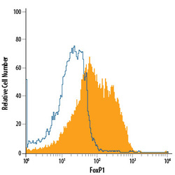
- Detection of FoxP1 in MCF-7 Human Cell Line by Flow Cytometry. MCF-7 human breast cancer cell line was stained with Mouse Anti-Human FoxP1 Monoclonal Antibody (Catalog # MAB45341, filled histogram) or isotype control antibody (Catalog # MAB002, open histogram), followed by Allophycocyanin-conjugated Anti-Mouse IgG Secondary Antibody (Catalog # F0101B). To facilitate intracellular staining, cells were fixed with 4% paraformaldehyde and permeabilized with methanol.