Explore
 Validate
 Learn
Learn Western blot
 Immunocytochemistry
Antibody data
- Antibody Data
- Antigen structure
- References [2]
- Comments [0]
- Validations
- Western blot [2]
- Immunohistochemistry [1]
- Flow cytometry [1]
Submit
Validation data
Reference
Comment
Report error
- Product number
- MAB45341 - Provider product page

- Provider
- Novus Biologicals
- Product name
- Mouse Monoclonal FoxP1 Antibody
- Antibody type
- Monoclonal
- Description
- Protein A or G purified from hybridoma culture supernatant. Detects human FoxP1 in direct ELISAs and Western blots.
- Reactivity
- Human
- Host
- Mouse
- Conjugate
- Unconjugated
- Isotype
- IgG
- Vial size
- 100 ug
- Concentration
- LYOPH
- Storage
- Use a manual defrost freezer and avoid repeated freeze-thaw cycles. 12 months from date of receipt, -20 to -70 degreesC as supplied. 1 month, 2 to 8 degreesC under sterile conditions after reconstitution. 6 months, -20 to -70 degreesC under sterile conditions after reconstitution.
Submitted references Cell cycle inhibitors protect motor neurons in an organoid model of Spinal Muscular Atrophy.
Regulated expression of the TPβ isoform of the human T prostanoid receptor by the tumour suppressors FOXP1 and NKX3.1: Implications for the role of thromboxane in prostate cancer.
Hor JH, Soh ES, Tan LY, Lim VJW, Santosa MM, Winanto, Ho BX, Fan Y, Soh BS, Ng SY
Cell death & disease 2018 Oct 27;9(11):1100
Cell death & disease 2018 Oct 27;9(11):1100
Regulated expression of the TPβ isoform of the human T prostanoid receptor by the tumour suppressors FOXP1 and NKX3.1: Implications for the role of thromboxane in prostate cancer.
O'Sullivan AG, Eivers SB, Mulvaney EP, Kinsella BT
Biochimica et biophysica acta. Molecular basis of disease 2017 Dec;1863(12):3153-3169
Biochimica et biophysica acta. Molecular basis of disease 2017 Dec;1863(12):3153-3169
No comments: Submit comment
Supportive validation
- Submitted by
- Novus Biologicals (provider)
- Main image
- Experimental details
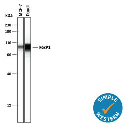
- Detection of Human FoxP1 by Simple WesternTM. Simple Western lane view shows lysates of MCF-7 human breast cancer cell line and Daudi human Burkitt's lymphoma cell line, loaded at 0.5 mg/mL. A specific band was detected for FoxP1 at approximately 99 kDa (as indicated) using 10 µg/mL of Mouse Anti-Human FoxP1 Monoclonal Antibody (Catalog # MAB45341). This experiment was conducted under reducing conditions and using the 12-230 kDa separation system.

- Submitted by
- Novus Biologicals (provider)
- Main image
- Experimental details
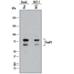
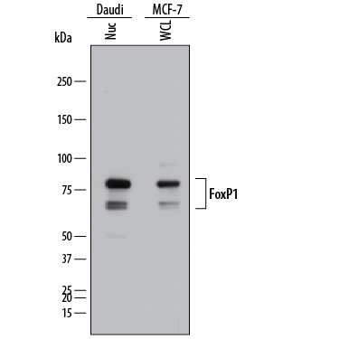
- Detection of Human FoxP1 by Western Blot. Western blot shows lysates of Daudi human Burkitt's lymphoma cell line and MCF-7 human breast cancer cell line. Gels were loaded with 25 µg of whole cell lysate (WCL) and 25 µg of nuclear extracts (Nuc). PVDF membrane was probed with 1 µg/mL of Mouse Anti-Human FoxP1 Monoclonal Antibody (Catalog # MAB45341) followed by HRP-conjugated Anti-Mouse IgG Secondary Antibody (Catalog # HAF018). Specific bands were detected for FoxP1 at approximately 65 and 80 kDa (as indicated). This experiment was conducted under reducing conditions and using Immunoblot Buffer Group 1.
Supportive validation
- Submitted by
- Novus Biologicals (provider)
- Main image

- Experimental details
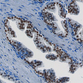
- FoxP1 in Human Prostate. FoxP1 was detected in immersion fixed paraffin-embedded sections of human prostate using Mouse Anti-Human FoxP1 Monoclonal Antibody (Catalog # MAB45341) at 15 µg/mL overnight at 4 °C. Before incubation with the primary antibody, tissue was subjected to heat-induced epitope retrieval using Antigen Retrieval Reagent-Basic (Catalog # CTS013). Tissue was stained using the Anti-Mouse HRP-DAB Cell & Tissue Staining Kit (brown; Catalog # CTS002) and counter-stained with hematoxylin (blue). Specific staining was localized to the nuclei of epithelial cells. View our protocol for Chromogenic IHC Staining of Paraffin-embedded Tissue Sections.
Supportive validation
- Submitted by
- Novus Biologicals (provider)
- Main image
- Experimental details
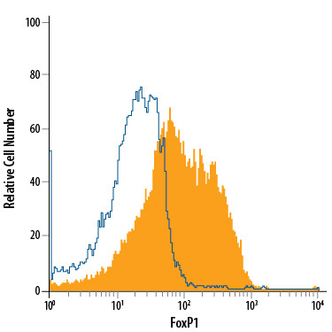
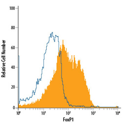
- Detection of FoxP1 in MCF-7 Human Cell Line by Flow Cytometry. MCF-7 human breast cancer cell line was stained with Mouse Anti-Human FoxP1 Monoclonal Antibody (Catalog # MAB45341, filled histogram) or isotype control antibody (Catalog # MAB002, open histogram), followed by Allophycocyanin-conjugated Anti-Mouse IgG Secondary Antibody (Catalog # F0101B). To facilitate intracellular staining, cells were fixed with 4% paraformaldehyde and permeabilized with methanol.