 Explore
 Validate
 Learn
Learn Western blot
Antibody data
- Antibody Data
- Antigen structure
- References [0]
- Comments [0]
- Validations
- Western blot [1]
- Immunohistochemistry [10]
- Blocking/Neutralizing [1]
Submit
Validation data
Reference
Comment
Report error
- Product number
- LS-C408138 - Provider product page

- Provider
- LSBio
- Product name
- CLOCK Antibody (aa75-109) LS-C408138
- Antibody type
- Polyclonal
- Description
- Immunogen affinity purified
- Reactivity
- Human, Mouse, Rat, Bovine, Canine, Chicken/Avian, Guinea Pig, Hamster, Horse, Rabbit, Sheep
- Host
- Rabbit
- Storage
- At -20°C for 1 year. After reconstitution, at 4°C for 1 month. It can also be aliquotted and stored frozen at -20°C for a longer time. Avoid freeze-thaw cycles.
No comments: Submit comment
Supportive validation
- Submitted by
- LSBio (provider)
- Main image
- Experimental details
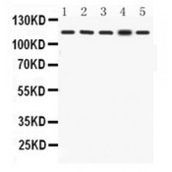
- KAT13D/CLOCK antibody Western blot. All lanes: Anti KAT13D/CLOCK at 0.5 ug/ml. Lane 1: Rat Skeletal Muscle Tissue Lysate at 50 ug. Lane 2: Mouse Skeletal Muscle Tissue Lysate at 50 ug. Lane 3: NIH3T3 Whole Cell Lysate at 40 ug. Lane 4: A549 Whole Cell Lysate at 40 ug. Lane 5: MCF7 Whole Cell Lysate at 40 ug. Predicted band size: 95 kD. Observed band size: 115 kD.

Supportive validation
- Submitted by
- LSBio (provider)
- Main image
- Experimental details
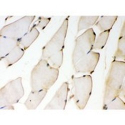
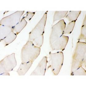
- KAT13D/CLOCK antibody IHC-paraffin. IHC(P): Mouse Skeletal Muscle Tissue.
- Submitted by
- LSBio (provider)
- Main image

- Experimental details
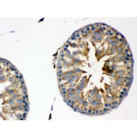
- KAT13D/CLOCK antibody IHC-paraffin. IHC(P): Rat Testis Tissue.

- Submitted by
- LSBio (provider)
- Main image
- Experimental details
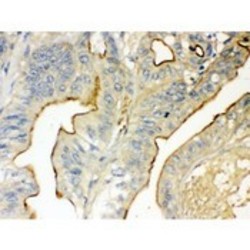
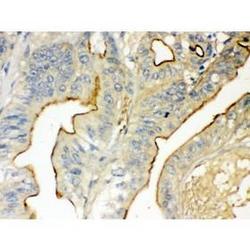
- KAT13D/CLOCK antibody IHC-paraffin. IHC(P): Human Intestinal Cancer Tissue.
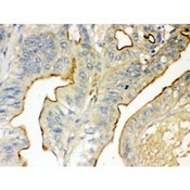
- Submitted by
- LSBio (provider)
- Main image

- Experimental details
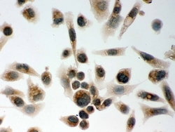
- IHC analysis of CLOCK using anti-CLOCK antibody. CLOCK was detected in immunocytochemical section of A549 cell. Heat mediated antigen retrieval was performed in citrate buffer (pH6, epitope retrieval solution) for 20 mins. The tissue section was blocked with 10% goat serum. The tissue section was then incubated with 1µg/ml rabbit anti-CLOCK Antibody overnight at 4°C. Biotinylated goat anti-rabbit IgG was used as secondary antibody and incubated for 30 minutes at 37°C. The tissue section was developed using Strepavidin-Biotin-Complex (SABC) with DAB as the chromogen.
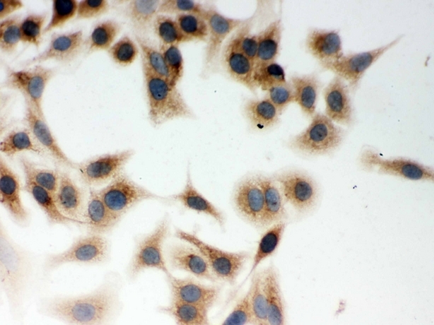
- Submitted by
- LSBio (provider)
- Main image
- Experimental details
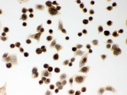
- IHC analysis of CLOCK using anti-CLOCK antibody. CLOCK was detected in immunocytochemical section of PC-3 cell. Heat mediated antigen retrieval was performed in citrate buffer (pH6, epitope retrieval solution) for 20 mins. The tissue section was blocked with 10% goat serum. The tissue section was then incubated with 1µg/ml rabbit anti-CLOCK Antibody overnight at 4°C. Biotinylated goat anti-rabbit IgG was used as secondary antibody and incubated for 30 minutes at 37°C. The tissue section was developed using Strepavidin-Biotin-Complex (SABC) with DAB as the chromogen.
- Submitted by
- LSBio (provider)
- Main image
- Experimental details
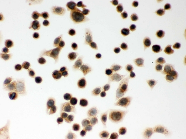
- IHC analysis of CLOCK using anti-CLOCK antibody. CLOCK was detected in immunocytochemical section of SW480 cell. Heat mediated antigen retrieval was performed in citrate buffer (pH6, epitope retrieval solution) for 20 mins. The tissue section was blocked with 10% goat serum. The tissue section was then incubated with 1µg/ml rabbit anti-CLOCK Antibody overnight at 4°C. Biotinylated goat anti-rabbit IgG was used as secondary antibody and incubated for 30 minutes at 37°C. The tissue section was developed using Strepavidin-Biotin-Complex (SABC) with DAB as the chromogen.

- Submitted by
- LSBio (provider)
- Main image
- Experimental details
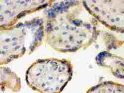
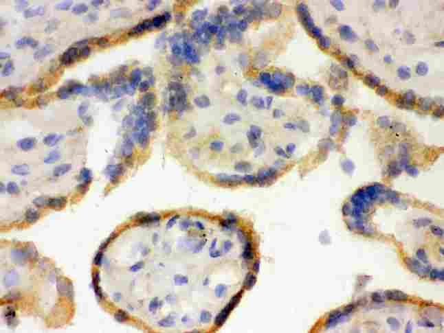
- IHC analysis of CLOCK using anti-CLOCK antibody. CLOCK was detected in frozen section of human placenta tissue . Heat mediated antigen retrieval was performed in citrate buffer (pH6, epitope retrieval solution) for 20 mins. The tissue section was blocked with 10% goat serum. The tissue section was then incubated with 1µg/ml rabbit anti-CLOCK Antibody overnight at 4°C. Biotinylated goat anti-rabbit IgG was used as secondary antibody and incubated for 30 minutes at 37°C. The tissue section was developed using Strepavidin-Biotin-Complex (SABC) with DAB as the chromogen.
- Submitted by
- LSBio (provider)
- Main image

- Experimental details
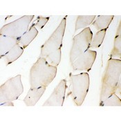
- KAT13D/CLOCK antibody IHC-paraffin. IHC(P): Mouse Skeletal Muscle Tissue.
- Submitted by
- LSBio (provider)
- Main image

- Experimental details
- KAT13D/CLOCK antibody IHC-paraffin. IHC(P): Rat Testis Tissue.
- Submitted by
- LSBio (provider)
- Main image
- Experimental details
- KAT13D/CLOCK antibody IHC-paraffin. IHC(P): Human Intestinal Cancer Tissue.

Supportive validation
- Submitted by
- LSBio (provider)
- Main image

- Experimental details
- KAT13D/CLOCK antibody Western blot. All lanes: Anti KAT13D/CLOCK at 0.5 ug/ml. Lane 1: Rat Skeletal Muscle Tissue Lysate at 50 ug. Lane 2: Mouse Skeletal Muscle Tissue Lysate at 50 ug. Lane 3: NIH3T3 Whole Cell Lysate at 40 ug. Lane 4: A549 Whole Cell Lysate at 40 ug. Lane 5: MCF7 Whole Cell Lysate at 40 ug. Predicted band size: 95 kD. Observed band size: 115 kD.
