Explore
 Validate
 Learn
Learn Western blot
Antibody data
- Antibody Data
- Antigen structure
- References [1]
- Comments [0]
- Validations
- Western blot [1]
- Immunohistochemistry [1]
- Other assay [8]
Submit
Validation data
Reference
Comment
Report error
- Product number
- PA5-41694 - Provider product page

- Provider
- Invitrogen Antibodies
- Product name
- DMRT2 Polyclonal Antibody
- Antibody type
- Polyclonal
- Antigen
- Synthetic peptide
- Description
- Peptide sequence: DPQAGSAAGD WEIDVESLEL EEDVCGAPRS TPPGPSPPPA DGDCEDDEDD Sequence homology: Human: 100%; Rabbit: 86%
- Reactivity
- Human
- Host
- Rabbit
- Isotype
- IgG
- Vial size
- 100 µL
- Concentration
- 1 mg/mL
- Storage
- -20° C, Avoid Freeze/Thaw Cycles
Submitted references DMRT2 Interacts With FXR and Improves Insulin Resistance in Adipocytes and a Mouse Model.
Tao J, Yu XL, Yuan YJ, Shen X, Liu J, Gu PP, Wang Z, Ma YT, Li GQ
Frontiers in endocrinology 2021;12:723623
Frontiers in endocrinology 2021;12:723623
No comments: Submit comment
Supportive validation
- Submitted by
- Invitrogen Antibodies (provider)
- Main image
- Experimental details
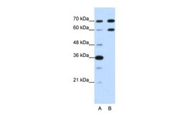
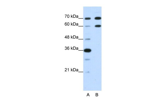
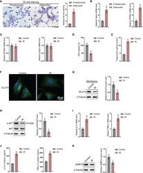
- Western blot analysis of human HepG2 cell lysate using an anti-DMRT2 polyclonal antibody (Product # PA5-41694).
Supportive validation
- Submitted by
- Invitrogen Antibodies (provider)
- Main image

- Experimental details
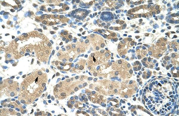
- Immunohistochemistry analysis of human kidney tissue using an anti-DMRT2 polyclonal antibody (Product # PA5-41694).
Supportive validation
- Submitted by
- Invitrogen Antibodies (provider)
- Main image
- Experimental details
- NULL
- Submitted by
- Invitrogen Antibodies (provider)
- Main image
- Experimental details
- NULL
- Submitted by
- Invitrogen Antibodies (provider)
- Main image
- Experimental details
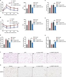
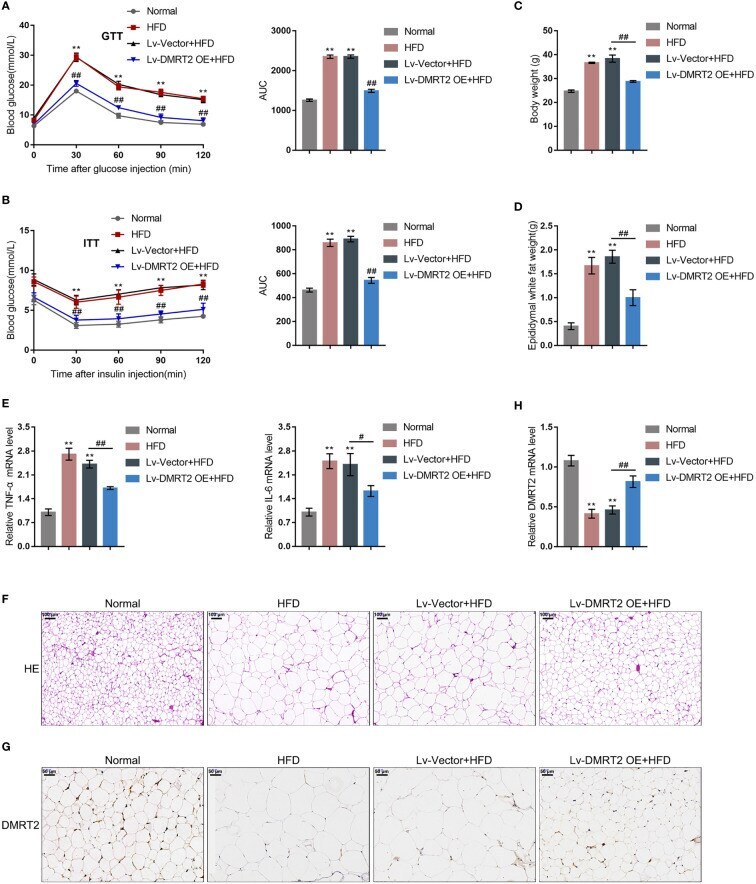
- In-vivo effects of DMRT2 in an insulin-resistant mouse model. The insulin-resistant model was established in mice as described. DMRT2 overexpression was achieved in control or model mice by injecting lentivirus containing DMRT2 OE. (A, B) Glucose tolerance test (GTT) and insulin tolerance test (ITT) were performed on mice as described and the area under curve (AUC) was calculated for each group. (C, D) Mice body weight and epididymal fat weight were examined. (E, H) The mRNA expression of TNF-alpha, IL-6, and DMRT2 in epididymal fat tissues was examined using qRT-PCR. (F) The histopathological characteristics of epididymal fat tissues were examined using H&E staining. (G) The levels of DMRT2 in epididymal fat tissues were examined using immunohistochemical (IHC) staining. ** P < 0.01, compared with the normal group; # P < 0.05, ## P < 0.01, compared Lv-Vector+HFD with the Lv-DMRT2 OE+HFD group.
- Submitted by
- Invitrogen Antibodies (provider)
- Main image
- Experimental details
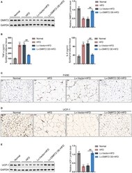
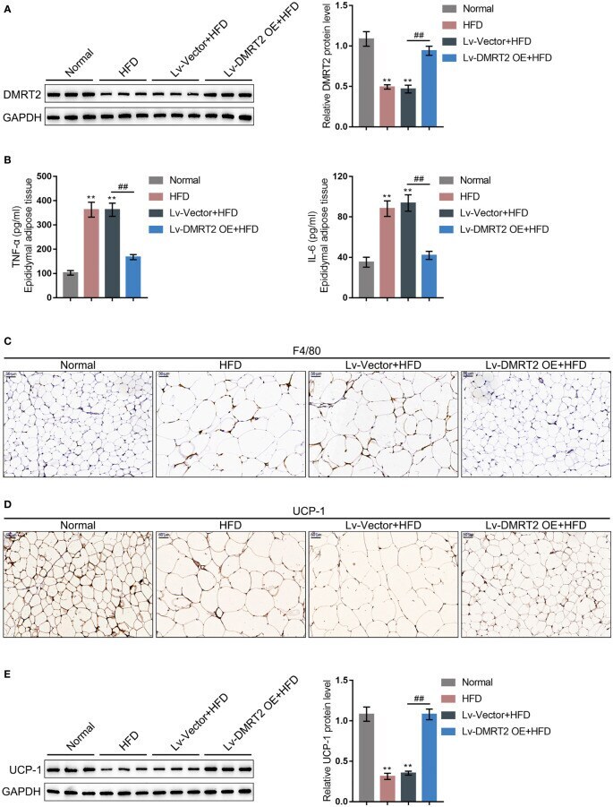
- Figure 2 DMRT2 reduced inflammation and increased white adipose browning in the insulin-resistant mouse model. (A) The protein level of DMRT2 in epididymal fat tissues was examined using immunoblotting. (B) The protein levels of TNF-alpha and IL-6 in epididymal adipose tissues were examined using ELISA. (C) The macrophage infiltration was determined using IHC staining for F4/80. (D, E) The brown adipocytes in epididymal adipose tissues were determined using IHC staining and immunoblotting for UCP-1. ** P < 0.01, compared with the normal group; ## P < 0.01, compared Lv-Vector+HFD with the Lv-DMRT2 OE+HFD group.
- Submitted by
- Invitrogen Antibodies (provider)
- Main image
- Experimental details
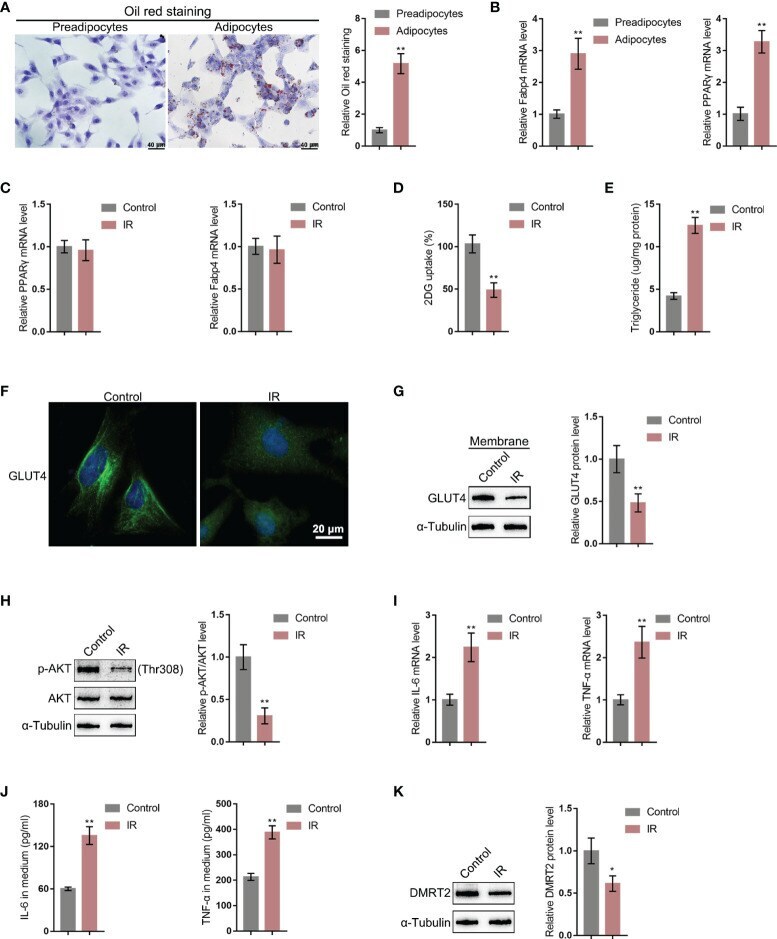
- Figure 3 DMRT2 is downregulated in insulin-resistant adipocyte model. (A) 3T3-L1 preadipocytes were induced toward adipogenesis and the formation of lip droplets in preadipocytes and adipocytes was confirmed using oil O red staining. (B) The mRNA expression levels of Fabp4 and PPARgamma in preadipocytes and adipocytes were examined using qRT-PCR. Then, the insulin-resistant adipocyte model was established in differentiated adipocytes as described and examined for Fabp4 and PPARgamma mRNA levels (C) , glucose uptake ability (D) , and triglyceride content (E) ; cellular membrane-located GLUT4 levels in control or insulin-resistant (IR) adipocytes by immunofluorescent (IF) staining (F) ; membrane protein levels of GLUT4 in control or IR adipocytes by immunoblotting (G) ; the protein levels of Akt and p-Akt in control or IR adipocytes by immunoblotting (H) ; the mRNA expression of TNF-alpha and IL-6 in control or IR adipocytes by qRT-PCR (I) ; the secretion levels of TNF-alpha and IL-6 in control or IR adipocytes by ELISA (J) ; and the protein levels of DMRT2 in control or IR adipocytes by immunoblotting (K) . * P < 0.05, ** P < 0.01.
- Submitted by
- Invitrogen Antibodies (provider)
- Main image
- Experimental details
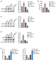
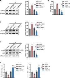
- In-vitro effects of DMRT2 on IR adipocytes. (A) DMRT2 overexpression or knockdown was achieved in adipocytes by transducing DMRT2-overexpressing vector (DMRT2 OE) or small interference RNA for DMRT2 (sh-DMRT2). The overexpression or knockdown of DMRT2 was confirmed using immunoblotting. Then, IR adipocytes were transfected with DMRT2 OE or sh-DMRT2 and examined for glucose uptake ability (B) ; protein levels of GLUT4 by immunoblotting (C) ; the protein levels of Akt and p-Akt by immunoblotting (D) ; and the mRNA expression of TNF-alpha and IL-6 in IR adipocytes by qRT-PCR (E) . * P < 0.05, ** P < 0.01, compared with the vector group; ## P < 0.01, compared sh-NC with the sh-DMRT2 group.

- Submitted by
- Invitrogen Antibodies (provider)
- Main image
- Experimental details
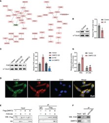
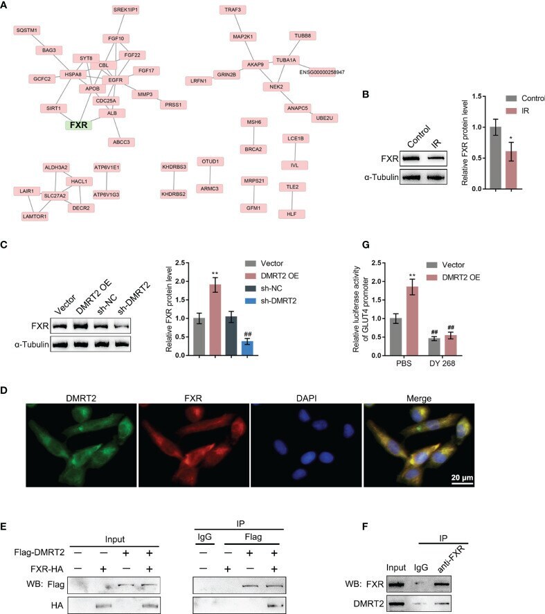
- Figure 5 DMRT2 directly interacts with FXR. (A) Protein-protein interaction analysis showing proteins that might interact with DMRT2. (B) The protein levels of FXR in IR adipocytes and IF adipocytes were examined using immunoblotting. (C) IR adipocytes were transfected with DMRT2 OE or sh-DMRT2 and examined for the protein levels of FXR by immunoblotting. (D) The interaction between DMRT2 and FXR was examined using IF staining. Green fluorescence indicated DMRT2. Red fluorescence indicated FXR. (E, F) The interaction between DMRT2 and FXR was examined using the co-IP assay. (G) 3T3-L1 cells were co-transfected with DMRT2 overexpression vector and GLUT4 promoter luciferase reporter vector. Then, the luciferase activity was measured. * P < 0.05, ** P < 0.01, compared with the control or vector group; ## P < 0.01, compared sh-NC with the sh-DMRT2 group.
- Submitted by
- Invitrogen Antibodies (provider)
- Main image
- Experimental details
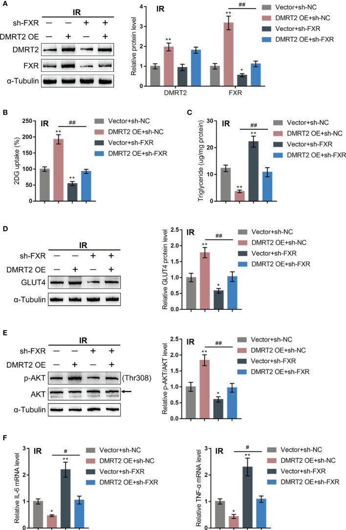
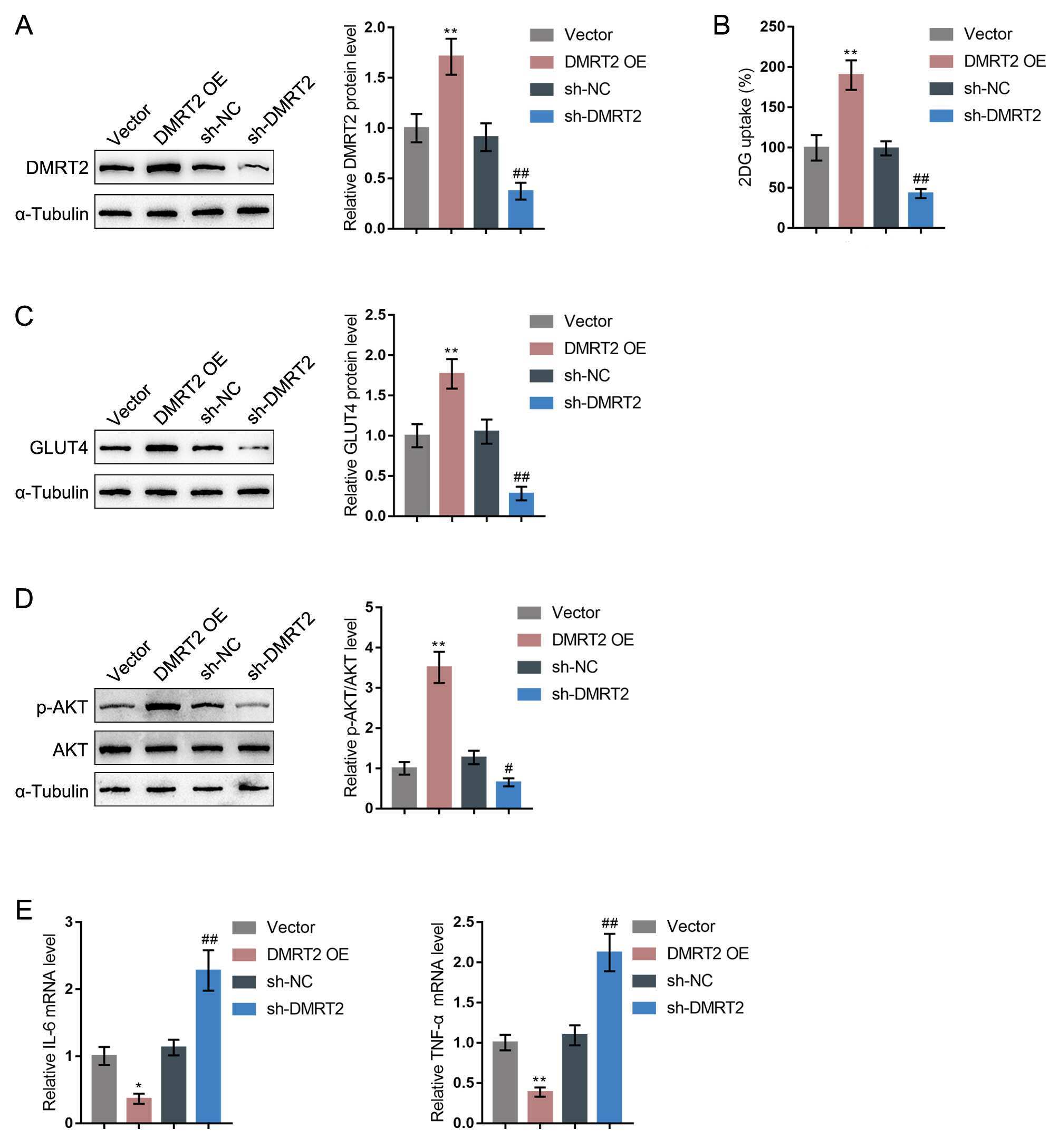
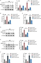
- Dynamic effects of DMRT2 and FXR on adipocyte resistance to insulin. IR adipocytes were co-transfected with DMRT2 OE and sh-FXR and examined for the protein levels of DMRT2 and FXR by immunoblotting (A) ; glucose uptake ability (B) , triglyceride content (C) , and the protein levels of GLUT4 by immunoblotting (D) ; the protein levels of Akt and p-Akt by immunoblotting (E) ; and the mRNA expression of TNF-alpha and IL-6 in control or IR adipocytes by qRT-PCR (F) . * P < 0.05, ** P < 0.01, compared with the Vector+sh-NC group; # P < 0.05, ## P < 0.01, compared Vector+sh-FXR with the DMRT2 OE+sh-FXR group.