Explore
 Validate
 Learn
Learn Western blot
 Immunohistochemistry
Antibody data
- Antibody Data
- Antigen structure
- References [0]
- Comments [0]
- Validations
- Western blot [1]
- Immunohistochemistry [6]
Submit
Validation data
Reference
Comment
Report error
- Product number
- AMAb90950 - Provider product page

- Provider
- Atlas Antibodies
- Proper citation
- Atlas Antibodies Cat#AMAb90950, RRID:AB_2665730
- Product name
- Anti-MYH6
- Antibody type
- Monoclonal
- Reactivity
- Human
- Host
- Mouse
- Conjugate
- Unconjugated
- Antigen sequence
QVEEDKVNSLSKSKVKLEQQVDDLEGSLEQEKKVR
MDLERAKRKLEGDLKLTQESIMDLENDKLQLEEKL
KKKEFDINQQNSKIEDEQVLALQLQKKLKENQARI
EELEEELEAERTARAKVEKLRSDLSRELEEISERL
EEA- Epitope
- Binds to an epitope located within the peptide sequence RELEEISERLEEA as determined by overlapping synthetic peptides.
- Isotype
- IgG
- Antibody clone number
- CL2162
- Vial size
- 100 µl
- Storage
- Store at +4°C for short term storage. Long time storage is recommended at -20°C.
No comments: Submit comment
Supportive validation
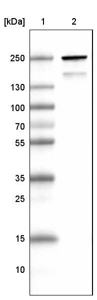
- Submitted by
- Atlas Antibodies (provider)
- Main image

- Experimental details
- Lane 1: Marker [kDa]Lane 2: Human skeletal muscle tissue lysate
Enhanced validation
Supportive validation
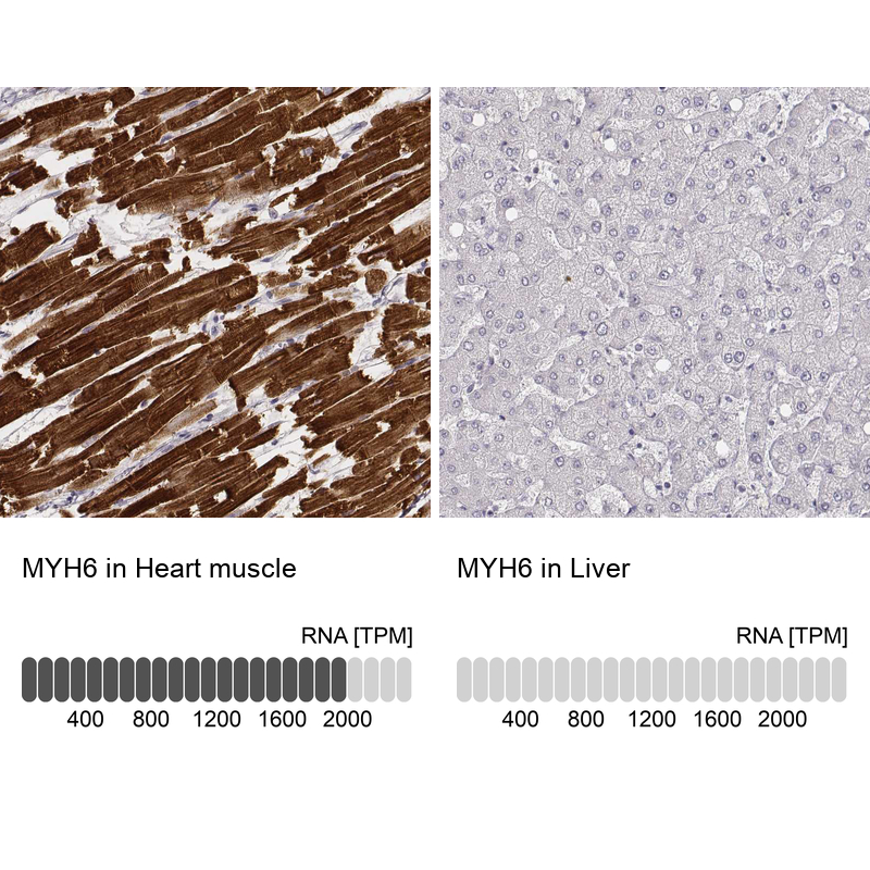
- Submitted by
- Atlas Antibodies (provider)
- Enhanced method
- Orthogonal validation
- Main image

- Experimental details
- Immunohistochemistry analysis in human heart muscle and liver tissues using AMAb90950 antibody. Corresponding MYH6 RNA-seq data are presented for the same tissues.
- Sample type
- HUMAN
Supportive validation
- Submitted by
- Atlas Antibodies (provider)
- Main image

- Experimental details
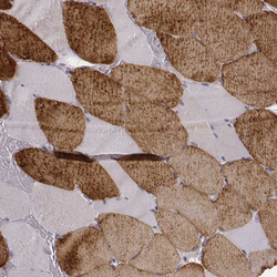
- Immunohistochemical staining of human skeletal muscle shows strong cytoplasmic immunoreactivity in a subset of striated muscle fibers.
- Submitted by
- Atlas Antibodies (provider)
- Main image
- Experimental details
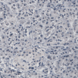
- Immunohistochemical staining of human pancreas shows absence of immunoreactivity (negative control).
- Submitted by
- Atlas Antibodies (provider)
- Main image

- Experimental details
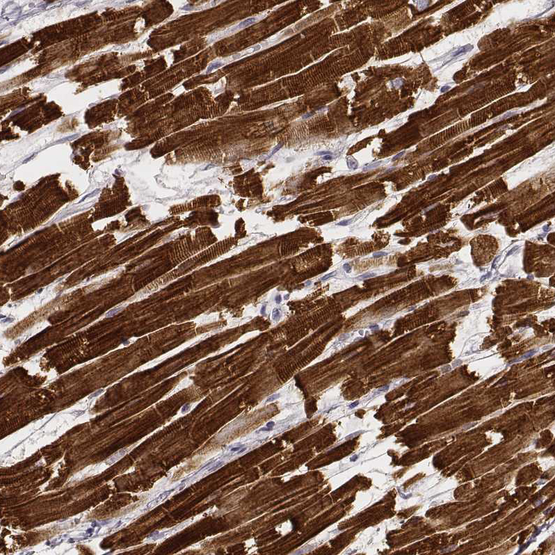
- Immunohistochemical staining of human heart muscle shows very strong cytoplasmic positivity in cardiomyocytes.
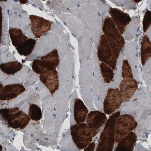
- Submitted by
- Atlas Antibodies (provider)
- Main image
- Experimental details
- Immunohistochemical staining of human skeletal muscle shows strong cytoplasmic positivity in striated muscle fibers.

- Submitted by
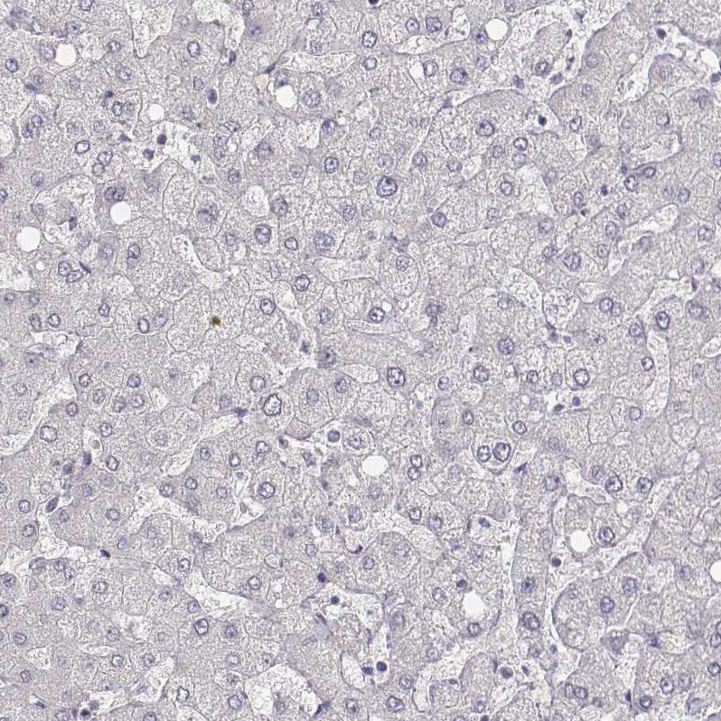
- Atlas Antibodies (provider)
- Main image

- Experimental details
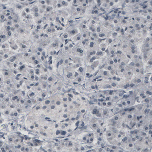
- Immunohistochemical staining of human liver shows no positivity in hepatocytes as expected.