Explore
 Validate
 Learn
Learn Immunohistochemistry
Antibody data
- Antibody Data
- Antigen structure
- References [7]
- Comments [0]
- Validations
- Immunohistochemistry [1]
Submit
Validation data
Reference
Comment
Report error
- Product number
- HPA001349 - Provider product page

- Provider
- Atlas Antibodies
- Proper citation
- Atlas Antibodies Cat#HPA001349, RRID:AB_1079437
- Product name
- Anti-MYH6
- Antibody type
- Polyclonal
- Description
- Polyclonal Antibody against Human MYH6, Gene description: myosin, heavy chain 6, cardiac muscle, alpha, Validated applications: IHC, Uniprot ID: P13533, Storage: Store at +4°C for short term storage. Long time storage is recommended at -20°C.
- Reactivity
- Human
- Host
- Rabbit
- Conjugate
- Unconjugated
- Isotype
- IgG
- Vial size
- 100 µl
- Concentration
- 0.1 mg/ml
- Storage
- Store at +4°C for short term storage. Long time storage is recommended at -20°C.
- Handling
- The antibody solution should be gently mixed before use.
Submitted references Expression of MyHC‐15 and ‐2x in human muscle spindles: An immunohistochemical study
Targeting the Wnt signaling pathway through R-spondin 3 identifies an anti-fibrosis treatment strategy for multiple organs
Study of the Expression Transition of Cardiac Myosin Using Polarization-Dependent SHG Microscopy
Letter to the editor: Comments on Stuart et al. (2016): “Myosin content of individual human muscle fibers isolated by laser capture microdissection”
Myosin content of individual human muscle fibers isolated by laser capture microdissection
Optimization of electrical stimulation parameters for cardiac tissue engineering
Dynamic expression patterns of leucine‐rich repeat containing protein 10 in the heart
Smerdu V
Journal of Anatomy 2023;243(5):826-841
Journal of Anatomy 2023;243(5):826-841
Targeting the Wnt signaling pathway through R-spondin 3 identifies an anti-fibrosis treatment strategy for multiple organs
Mukhopadhyay P, Zhang M, Haughey M, Wang N, Blease K, Kapoun A, Couto S, Belka I, Hoey T, Groza M, Hartke J, Bennett B, Cain J, Gurney A, Benish B, Castiglioni P, Drew C, Lachowicz J, Carayannopoulos L, Nathan S, Distler J, Brenner D, Hariharan K, Cho H, Xie W
PLOS ONE 2020;15(3):e0229445
PLOS ONE 2020;15(3):e0229445
Study of the Expression Transition of Cardiac Myosin Using Polarization-Dependent SHG Microscopy
Yuan C, Zhao X, Wang Z, Borg T, Ye T, Khalpey Z, Runyan R, Shao Y, Gao B
Biophysical Journal 2020;118(5):1058-1066
Biophysical Journal 2020;118(5):1058-1066
Letter to the editor: Comments on Stuart et al. (2016): “Myosin content of individual human muscle fibers isolated by laser capture microdissection”
Schiaffino S, Murgia M, Leinwand L, Reggiani C
American Journal of Physiology-Cell Physiology 2016;311(6):C1048-C1049
American Journal of Physiology-Cell Physiology 2016;311(6):C1048-C1049
Myosin content of individual human muscle fibers isolated by laser capture microdissection
Stuart C, Stone W, Howell M, Brannon M, Hall H, Gibson A, Stone M
American Journal of Physiology-Cell Physiology 2016;310(5):C381-C389
American Journal of Physiology-Cell Physiology 2016;310(5):C381-C389
Optimization of electrical stimulation parameters for cardiac tissue engineering
Tandon N, Marsano A, Maidhof R, Wan L, Park H, Vunjak-Novakovic G
Journal of Tissue Engineering and Regenerative Medicine 2011;5(6):e115-e125
Journal of Tissue Engineering and Regenerative Medicine 2011;5(6):e115-e125
Dynamic expression patterns of leucine‐rich repeat containing protein 10 in the heart
Kim K, Kim T, Micales B, Lyons G, Lee Y
Developmental Dynamics 2007;236(8):2225-2234
Developmental Dynamics 2007;236(8):2225-2234
No comments: Submit comment
Supportive validation
- Submitted by
- Atlas Antibodies (provider)
- Enhanced method
- Orthogonal validation
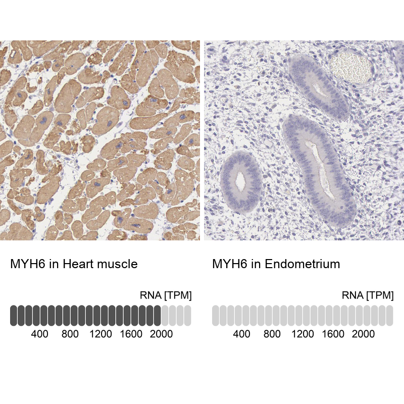
- Main image
- Experimental details
- Immunohistochemistry analysis in human heart muscle and endometrium tissues using HPA001349 antibody. Corresponding MYH6 RNA-seq data are presented for the same tissues.
- Sample type
- Human
- Protocol
- Protocol