Explore
 Validate
 Learn
Learn Western blot
Antibody data
- Antibody Data
- Antigen structure
- References [1]
- Comments [0]
- Validations
- Western blot [4]
- Immunocytochemistry [1]
- Immunohistochemistry [1]
Submit
Validation data
Reference
Comment
Report error
- Product number
- GTX105789 - Provider product page

- Provider
- GeneTex
- Proper citation
- GeneTex Cat#GTX105789, RRID:AB_10721688
- Product name
- L-Plastin antibody
- Antibody type
- Polyclonal
- Reactivity
- Human, Mouse
- Host
- Rabbit
Submitted references High glucose-induced proteome alterations in hepatocytes and its possible relevance to diabetic liver disease.
Chen JY, Chou HC, Chen YH, Chan HL
The Journal of nutritional biochemistry 2013 Nov;24(11):1889-910
The Journal of nutritional biochemistry 2013 Nov;24(11):1889-910
No comments: Submit comment
Supportive validation
- Submitted by
- GeneTex (provider)
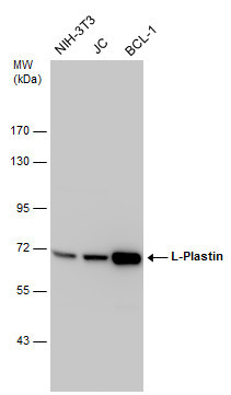
- Main image
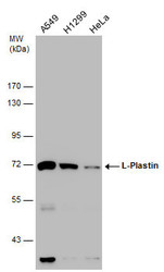
- Experimental details
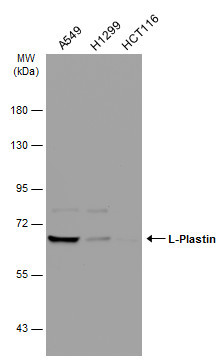
- Various whole cell extracts (30 ?g) were separated by 7.5% SDS-PAGE, and the membrane was blotted with L-Plastin antibody (GTX105789) diluted at 1:10000.
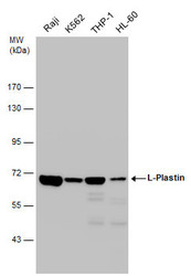
- Submitted by
- GeneTex (provider)
- Main image
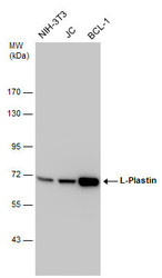
- Experimental details
- Various whole cell extracts (30 ?g) were separated by 7.5% SDS-PAGE, and the membrane was blotted with L-Plastin antibody (GTX105789) diluted at 1:10000.
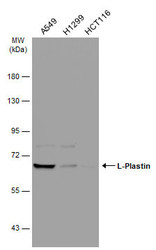
- Submitted by
- GeneTex (provider)
- Main image
- Experimental details
- Various whole cell extracts (30 ?g) were separated by 7.5% SDS-PAGE, and the membrane was blotted with L-Plastin antibody (GTX105789) diluted at 1:10000.
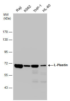
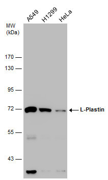
- Submitted by
- GeneTex (provider)
- Main image
- Experimental details
- Various whole cell extracts (30 ?g) were separated by 7.5% SDS-PAGE, and the membrane was blotted with L-Plastin antibody (GTX105789) diluted at 1:10000. The HRP-conjugated anti-rabbit IgG antibody (GTX213110-01) was used to detect the primary antibody.
Supportive validation
- Submitted by
- GeneTex (provider)
- Main image
- Experimental details
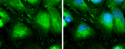
- L-Plastin antibody detects L-Plastin protein at cytoplasm and focal adhesion site by immunofluorescent analysis.Sample: A431 cells were fixed in ice-cold MeOH for 5 min.Green: L-Plastin protein stained by L-Plastin antibody (GTX105789) diluted at 1:500.Blue: Hoechst 33342 staining.

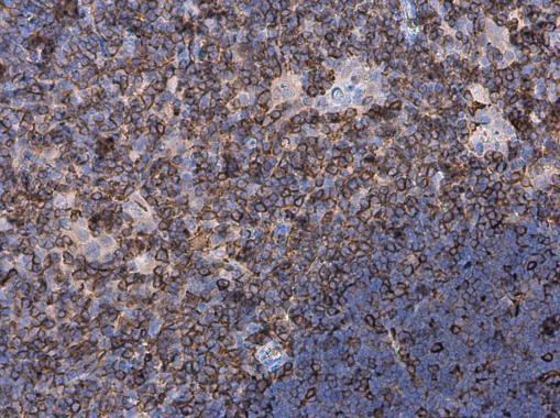
Supportive validation
- Submitted by
- GeneTex (provider)
- Main image
- Experimental details
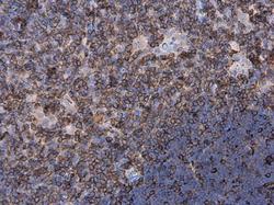
- Plastin L antibody detects Plastin L protein at cell membrane and cytoplasm in mouse thymus gland by immunohistochemical analysis. Sample: Paraffin-embedded mouse thymus gland. Plastin L antibody (GTX105789) diluted at 1:500.