 Explore
 Validate
 Learn
Learn Western blot
 Immunocytochemistry
 Immunohistochemistry
Antibody data
- Antibody Data
- Antigen structure
- References [1]
- Comments [0]
- Validations
- Immunocytochemistry [1]
- Chromatin Immunoprecipitation [2]
Submit
Validation data
Reference
Comment
Report error
- Product number
- MA1-12421 - Provider product page

- Provider
- Invitrogen Antibodies
- Product name
- LEF1 Monoclonal Antibody (REMB6)
- Antibody type
- Monoclonal
- Antigen
- Recombinant full-length protein
- Description
- A suggested positive control for this product is Jurkat cell lysate.
- Reactivity
- Human, Mouse
- Host
- Mouse
- Isotype
- IgG
- Antibody clone number
- REMB6
- Vial size
- 100 μg
- Concentration
- 1 mg/mL
- Storage
- -20°C, Avoid Freeze/Thaw Cycles
Submitted references Association of nuclear-localized Nemo-like kinase with heat-shock protein 27 inhibits apoptosis in human breast cancer cells.
Shaw-Hallgren G, Chmielarska Masoumi K, Zarrizi R, Hellman U, Karlsson P, Helou K, Massoumi R
PloS one 2014;9(5):e96506
PloS one 2014;9(5):e96506
No comments: Submit comment
Supportive validation
- Submitted by
- Invitrogen Antibodies (provider)
- Main image

- Experimental details
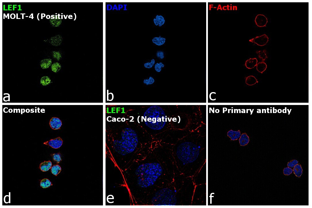
- Immunofluorescence analysis of LEF1 was performed using 70% confluent log phase MOLT-4 cells. The cells were fixed with 4% paraformaldehyde for 10 minutes, permeabilized with 0.1% Triton™ X-100 for 15 minutes, and blocked with 2% BSA for 45 minutes at room temperature. The cells were labeled with LEF1 Monoclonal Antibody (REMB6) (Product # MA1-12421) at 1:200 dilution in 0.1% BSA, incubated at 4 degree celsius overnight and then labeled with Goat anti-Mouse IgG (H+L) Highly Cross-Adsorbed Secondary Antibody, Alexa Fluor Plus 488 (Product # A32723), (1:3000 dilution), for 45 minutes at room temperature (Panel a: Green). Nuclei (Panel b: Blue) were stained with ProLong™ Diamond Antifade Mountant with DAPI (Product # P36962). F-actin (Panel c: Red) was stained with Rhodamine Phalloidin (Product # R415, 1:300). Panel d represents the merged image showing nucleus localization. Panel e represents merged image for Caco-2 cells showing no staining for LEF-1. Panel f represents control cells with no primary antibody to assess background. The images were captured at 60X magnification.
Supportive validation
- Submitted by
- Invitrogen Antibodies (provider)
- Main image

- Experimental details
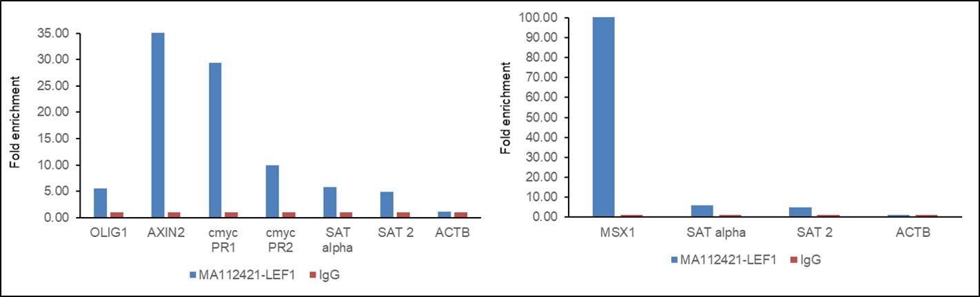
- Chromatin Immunoprecipitation (ChIP) assay of endogenous LEF1 protein using LEF1 Antibody: ChIP was performed using LEF1 Monoclonal Antibody (REMB6) (Product # MA1-12421, 5 µg) on sheared chromatin from HCT 116 cells using the MAGnify ChIP System kit (Product # 49-2024). Normal Mouse IgG was used as a negative IP control. The purified DNA was analyzed by qPCR using primers binding to OLIG1, AXIN2, c myc promoter (region1- PR1), c myc promoter (region2- PR2) and MSX1 (active) and SAT alpha, SAT2 satellite repeats and Actin beta (Inactive). Data is presented as fold enrichment of the antibody signal versus the negative control IgG using the comparative CT method.
- Submitted by
- Invitrogen Antibodies (provider)
- Main image

- Experimental details
- Chromatin Immunoprecipitation (ChIP) assay of endogenous LEF1 protein using LEF1 Antibody: ChIP was performed using LEF1 Monoclonal Antibody (REMB6) (Product # MA1-12421, 5 µg) on sheared chromatin from HCT 116 cells using the MAGnify ChIP System kit (Product # 49-2024). Normal Mouse IgG was used as a negative IP control. The purified DNA was analyzed by qPCR using primers binding to OLIG1, AXIN2, c myc promoter (region1- PR1), c myc promoter (region2- PR2) and MSX1 (active) and SAT alpha, SAT2 satellite repeats and Actin beta (Inactive). Data is presented as fold enrichment of the antibody signal versus the negative control IgG using the comparative CT method.
