Explore
 Validate
 Learn
Learn Western blot
Antibody data
- Antibody Data
- Antigen structure
- References [1]
- Comments [0]
- Validations
- Western blot [2]
- Immunocytochemistry [1]
- Immunohistochemistry [1]
Submit
Validation data
Reference
Comment
Report error
- Product number
- AF7197 - Provider product page

- Provider
- R&D Systems
- Product name
- Human/Mouse Lipoprotein Lipase/LPL Antibody
- Antibody type
- Polyclonal
- Description
- Antigen Affinity-purified. Detects human and mouse Lipoprotein Lipase/LPL in Western blots. Detects human Lipoprotein Lipase/LPL in direct ELISAs and less than 1% cross-reactivity with recombinant human (rh) LIPG, rhLIPI, and rhPNLIPRP1 is observed.
- Reactivity
- Human, Mouse
- Host
- Goat
- Conjugate
- Unconjugated
- Antigen sequence
P06858- Isotype
- IgG
- Vial size
- 100 ug
- Concentration
- LYOPH
- Storage
- Use a manual defrost freezer and avoid repeated freeze-thaw cycles. 12 months from date of receipt, -20 to -70 °C as supplied. 1 month, 2 to 8 °C under sterile conditions after reconstitution. 6 months, -20 to -70 °C under sterile conditions after reconstitution.
Submitted references Firemaster® 550 and its components isopropylated triphenyl phosphate and triphenyl phosphate enhance adipogenesis and transcriptional activity of peroxisome proliferator activated receptor (Pparγ) on the adipocyte protein 2 (aP2) promoter.
Tung EWY, Ahmed S, Peshdary V, Atlas E
PloS one 2017;12(4):e0175855
PloS one 2017;12(4):e0175855
No comments: Submit comment
Supportive validation
- Submitted by
- R&D Systems (provider)
- Main image
- Experimental details
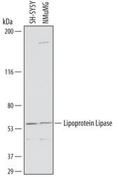
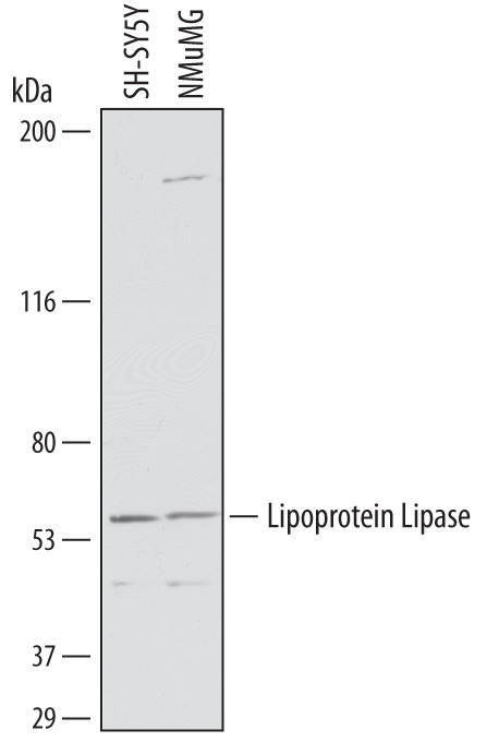
- Detection of Human and Mouse Lipoprotein Lipase/LPL by Western Blot. Western blot shows lysates of SH-SY5Y human neuroblastoma cell line and NMuMG mouse mammary gland epithelial cell line. PVDF membrane was probed with 1 µg/mL of Goat Anti-Human Lipoprotein Lipase/LPL Antigen Affinity-purified Polyclonal Antibody (Catalog # AF7197) followed by HRP-conjugated Anti-Goat IgG Secondary Antibody (Catalog # HAF019). A specific band was detected for Lipoprotein Lipase/LPL at approximately 56 kDa (as indicated). This experiment was conducted under reducing conditions and using Immunoblot Buffer Group 1.
- Submitted by
- R&D Systems (provider)
- Main image
- Experimental details
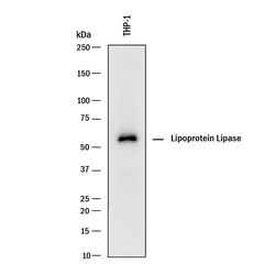
- Detection of Human Lipoprotein Lipase/LPL by Western Blot. Western blot shows lysates of THP-1 human acute monocytic leukemia cell line. PVDF membrane was probed with 1 µg/mL of Goat Anti-Human/Mouse Lipoprotein Lipase/LPL Antigen Affinity-purified Polyclonal Antibody (Catalog # AF7197) followed by HRP-conjugated Anti-Goat IgG Secondary Antibody (Catalog # HAF017). A specific band was detected for Lipoprotein Lipase/LPL at approximately 55 kDa (as indicated). This experiment was conducted under reducing conditions and using Immunoblot Buffer Group 1.

Supportive validation
- Submitted by
- R&D Systems (provider)
- Main image

- Experimental details
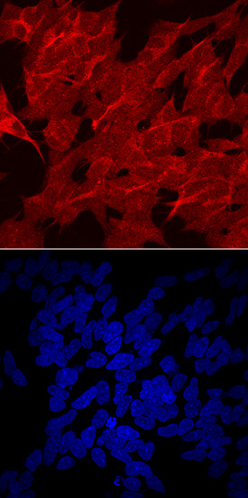
- Lipoprotein Lipase/LPL in SH-SY5Y Human Cell Line. Lipoprotein Lipase/LPL was detected in immersion fixed SH-SY5Y human neuroblastoma cell line using Goat Anti-Human Lipoprotein Lipase/LPL Antigen Affinity-purified Polyclonal Antibody (Catalog # AF7197) at 10 µg/mL for 3 hours at room temperature. Cells were stained using the NorthernLights™ 557-conjugated Anti-Goat IgG Secondary Antibody (red, upper panel; Catalog # NL001) and counterstained with DAPI (blue, lower panel). Specific staining was localized to cell surfaces and cytoplasm. View our protocol for Fluorescent ICC Staining of Cells on Coverslips. This application has not been tested in mouse samples.
Supportive validation
- Submitted by
- R&D Systems (provider)
- Main image

- Experimental details
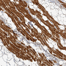
- Lipoprotein Lipase/LPL in Human Heart. Lipoprotein Lipase/LPL was detected in immersion fixed paraffin-embedded sections of human heart using Goat Anti-Human/Mouse Lipoprotein Lipase/LPL Antigen Affinity-purified Polyclonal Antibody (Catalog # AF7197) at 3 µg/mL for 1 hour at room temperature followed by incubation with the Anti-Goat IgG VisUCyte™ HRP Polymer Antibody (Catalog # VC004). Tissue was stained using DAB (brown) and counterstained with hematoxylin (blue). Specific staining was localized to cardiomyocytes. View our protocol for IHC Staining with VisUCyte HRP Polymer Detection Reagents.