Explore
 Validate
 Learn
Learn Western blot
Antibody data
- Antibody Data
- Antigen structure
- References [1]
- Comments [0]
- Validations
- Western blot [5]
- Immunocytochemistry [2]
- Chromatin Immunoprecipitation [1]
Submit
Validation data
Reference
Comment
Report error
- Product number
- 702016 - Provider product page

- Provider
- Invitrogen Antibodies
- Product name
- SOX9 Recombinant Rabbit Monoclonal Antibody (7H13L8)
- Antibody type
- Monoclonal
- Antigen
- Synthetic peptide
- Description
- This antibody is predicted to react with Monkey, Pig, Mouse
- Antibody clone number
- 7H13L8
- Concentration
- 0.5 mg/mL
Submitted references PLK2 modulation of enriched TAp73 affects osteogenic differentiation and prognosis in human osteosarcoma.
Li W, Zhang X, Xi X, Li Y, Quan H, Liu S, Wu L, Wu P, Lan W, Shao Y, Li H, Chen K, Hu Z
Cancer medicine 2020 Jun;9(12):4371-4385
Cancer medicine 2020 Jun;9(12):4371-4385
No comments: Submit comment
Supportive validation
- Submitted by
- Invitrogen Antibodies (provider)
- Main image

- Experimental details
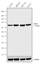
- Western blot analysis was performed on Modified Whole cell extracts (1% SDS) (30 µg lysate) of C2C12 (Lane 1), NIH/3T3 (Lane 2), HCT 116 (Lane 3) and Caco-2 (Lane 4). The blots were probed with Anti-SOX9 Recombinant Rabbit Monoclonal Antibody (Product # 702016, 2.5 µg/mL) and detected by chemiluminescence using Goat anti-Rabbit IgG (H+L) Superclonal™ Secondary Antibody, HRP conjugate (Product # A27036, 0.25 µg/mL, 1:4000 dilution). A 70 kDa band corresponding to SOX9 was observed across the cell lines tested. Known quantity of protein samples were electrophoresed using Novex®NuPAGE®4-12% Bis-Tris gel (Product # NP0321BOX), XCell SureLock™ Electrophoresis System (Product # EI0002) and Novex® Sharp Pre-Stained Protein Standard (Product # LC5800). Resolved proteins were then transferred onto a nitrocellulose membrane with iBlot® Dry Blotting System (Product # IB21001). The membrane was probed with the relevant primary and secondary Antibody following blocking with 5% skimmed milk. Chemiluminescent detection was performed using Pierce™ ECL Western blotting Substrate (Product # 32106).

- Submitted by
- Invitrogen Antibodies (provider)
- Main image

- Experimental details
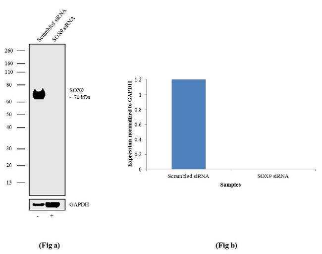
- Knockdown of SOX9 was achieved by transfecting PC-3 cells with SOX9 specific validated siRNA (Silencer® select Product # s13308). Western blot analysis (Fig a) was performed using Modified Whole cell extracts (1% SDS) from the SOX9 knock down cells (Lane 2) and non-specific scrambled siRNA transfected cells (Lane 1). The blots were probed with Anti-SOX9 Recombinant Rabbit Monoclonal Antibody (Product # 702016, 1-3 µg/mL) and Goat anti-Rabbit IgG (H+L) Superclonal™ Secondary Antibody, HRP conjugate (Product # A27036, 0.25 µg/mL, 1:4000 dilution). Densitometric analysis of this Western blot is shown in histogram (Fig b). Loss of signal upon siRNA mediated knock down confirms that antibody is specific to SOX9.
- Submitted by
- Invitrogen Antibodies (provider)
- Main image
- Experimental details
- Western blot analysis was performed on Modified Whole cell extracts (1% SDS) (30 µg lysate) of C2C12 (Lane 1), NIH/3T3 (Lane 2), HCT 116 (Lane 3) and Caco-2 (Lane 4). The blots were probed with Anti-SOX9 Recombinant Rabbit Monoclonal Antibody (Product # 702016, 2.5 µg/mL) and detected by chemiluminescence using Goat anti-Rabbit IgG (H+L) Superclonal™ Secondary Antibody, HRP conjugate (Product # A27036, 0.25 µg/mL, 1:4000 dilution). A 70 kDa band corresponding to SOX9 was observed across the cell lines tested. Known quantity of protein samples were electrophoresed using Novex®NuPAGE®4-12% Bis-Tris gel (Product # NP0321BOX), XCell SureLock™ Electrophoresis System (Product # EI0002) and Novex® Sharp Pre-Stained Protein Standard (Product # LC5800). Resolved proteins were then transferred onto a nitrocellulose membrane with iBlot® Dry Blotting System (Product # IB21001). The membrane was probed with the relevant primary and secondary Antibody following blocking with 5% skimmed milk. Chemiluminescent detection was performed using Pierce™ ECL Western blotting Substrate (Product # 32106).

- Submitted by
- Invitrogen Antibodies (provider)
- Main image

- Experimental details
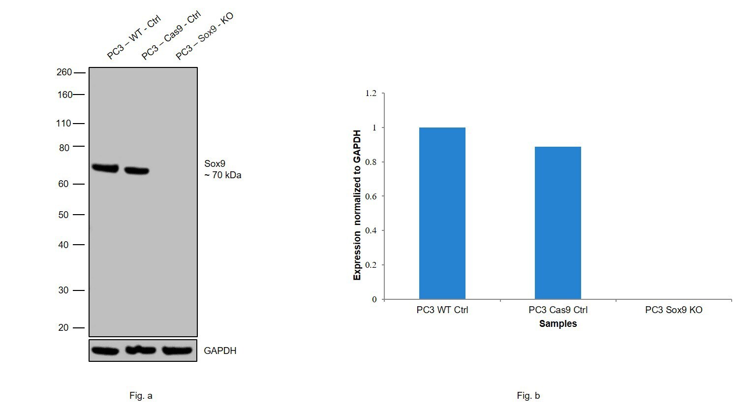
- Western blot analysis of SOX9 was performed by loading 20 µg of PC3 wild type (Lane 1), PC3 Cas9 control (Lane 2), PC3 SOX9 knockout (Lane 3) whole cell extracts. The blot was probed with Anti-SOX9 Recombinant Rabbit Monoclonal Antibody (7H13L8) (Product # 702016) (2 µg/mL) and Goat anti-Rabbit IgG (H+L), Superclonal™ Recombinant Secondary Antibody, HRP (Product # A27036) (1:4000 dilution). Loss of signal upon CRISPR mediated knockout (KO) confirms that antibody is specific to SOX9.
Supportive validation
- Submitted by
- Invitrogen Antibodies (provider)
- Main image

- Experimental details
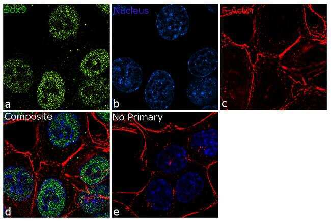
- For immunofluorescence analysis, HCT116 cells were fixed and permeabilized for detection of endogenous SOX9 using Anti- SOX9 Recombinant Rabbit Monoclonal Antibody (Product # 702016, 5 µg/mL) and labeled with Goat anti-Rabbit IgG (H+L) Superclonal™ Secondary Antibody, Alexa Fluor® 488 conjugate (Product # A27034, 1:2000). Panel a) shows representative cells that were stained for detection and localization of SOX9 protein (green), Panel b) is stained for nuclei (blue) using SlowFade® Gold Antifade Mountant with DAPI (Product # S36938). Panel c) represents cytoskeletal F-actin staining using Rhodamine Phalloidin (Product # R415, 1:300). Panel d) is a composite image of Panels a, b and c clearly demonstrating nuclear localization of SOX9. Panel e) represents control cells with no primary antibody to assess background. The images were captured at 60X magnification.
- Submitted by
- Invitrogen Antibodies (provider)
- Main image
- Experimental details
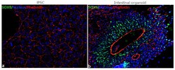
- For immunofluorescence analysis, iPSC differentiated to Intestinal organoids were fixed and permeabilized for detection of endogenous SOX9 using Anti-SOX9 Recombinant Rabbit monoclonal Antibody (Product # 702016, 1:100 dilution) and labeled with Goat anti-Rabbit IgG (H+L) Superclonal™ Secondary Antibody, Alexa Fluor® 488 conjugate (Product # A27034, 1:2000). Panel b) shows representative cells that were stained for detection and localization of SOX9 protein (green) in the nucleus of cells lining the lumen of intestinal organoids in comparison to iPSC (a).

Supportive validation
- Submitted by
- Invitrogen Antibodies (provider)
- Main image

- Experimental details
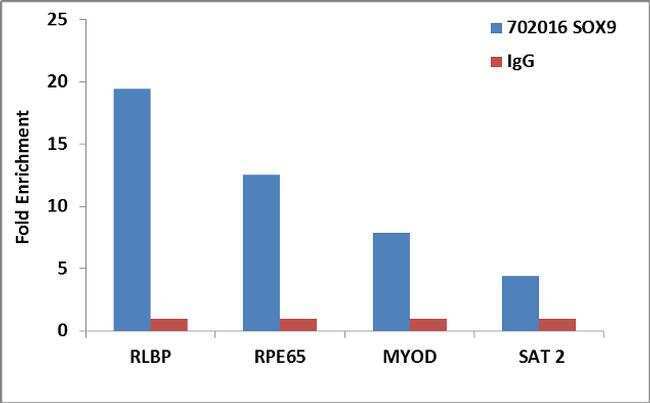
- Enrichment of endogenous SOX9 protein at specific gene loci using Anti-SOX9 Recombinant Rabbit Monoclonal Antibody: Chromatin Immunoprecipitation (ChIP) was performed using Anti-SOX9 Recombinant Rabbit Monoclonal Antibody (Product # 702016, 5 µg) on sheared chromatin from 2 million HCT 116 cells using the MAGnify ChIP system kit (Product # 49-2024). Normal Rabbit IgG (1 µg) was used as a negative IP control. The purified DNA was analyzed by 7500 Fast qPCR system (Product # 4351106) with optimized PCR primer pairs for the promoters of the RLBP, RPE65 used as positive control target genes, and the promoter region of the MYOD, SAT2 satellite repeat used as negative control target gene. Data is presented as fold enrichment of the antibody signal versus the negative control IgG using the comparative CT method.