 Explore
 Validate
 Learn
Learn Immunocytochemistry
 Immunohistochemistry
Antibody data
- Antibody Data
- Antigen structure
- References [2]
- Comments [0]
- Validations
- Immunocytochemistry [1]
- Chromatin Immunoprecipitation [1]
Submit
Validation data
Reference
Comment
Report error
- Product number
- HPA003256 - Provider product page

- Provider
- Atlas Antibodies
- Proper citation
- Atlas Antibodies Cat#HPA003256, RRID:AB_1079356
- Product name
- Anti-MEIS2
- Antibody type
- Polyclonal
- Description
- Polyclonal Antibody against Human MEIS2, Gene description: Meis homeobox 2, Alternative Gene Names: HsT18361, MRG1, Validated applications: IHC, ICC, ChIP, Uniprot ID: O14770, Storage: Store at +4°C for short term storage. Long time storage is recommended at -20°C.
- Reactivity
- Human
- Host
- Rabbit
- Conjugate
- Unconjugated
- Isotype
- IgG
- Vial size
- 100 µl
- Concentration
- 0.4 mg/ml
- Storage
- Store at +4°C for short term storage. Long time storage is recommended at -20°C.
- Handling
- The antibody solution should be gently mixed before use.
Submitted references MEIS2 Is an Adrenergic Core Regulatory Transcription Factor Involved in Early Initiation of TH-MYCN-Driven Neuroblastoma Formation
Lmx1b and FoxC Combinatorially Regulate Podocin Expression in Podocytes
De Wyn J, Zimmerman M, Weichert-Leahey N, Nunes C, Cheung B, Abraham B, Beckers A, Volders P, Decaesteker B, Carter D, Look A, De Preter K, Van Loocke W, Marshall G, Durbin A, Speleman F, Durinck K
Cancers 2021;13(19):4783
Cancers 2021;13(19):4783
Lmx1b and FoxC Combinatorially Regulate Podocin Expression in Podocytes
He B, Ebarasi L, Zhao Z, Guo J, Ojala J, Hultenby K, De Val S, Betsholtz C, Tryggvason K
Journal of the American Society of Nephrology 2014;25(12):2764-2777
Journal of the American Society of Nephrology 2014;25(12):2764-2777
No comments: Submit comment
Supportive validation
- Submitted by
- Atlas Antibodies (provider)
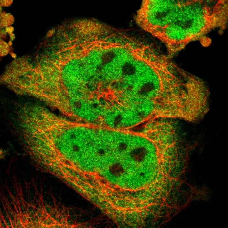
- Main image

- Experimental details
- Immunofluorescent staining of human cell line A-431 shows localization to nucleoplasm & cytosol.
- Sample type
- Human
Supportive validation
- Submitted by
- Atlas Antibodies (provider)
- Main image
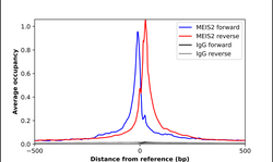
- Experimental details
- ChIP-Exo-Seq composite graph for Anti-MEIS2 (HPA003256, Lot A118643) tested in K562 cells. Strand-specific reads (blue: forward, red: reverse) and IgG controls (black: forward, grey: reverse) are plotted against the distance from a composite set of reference binding sites. The antibody exhibits robust target enrichment compared to a non-specific IgG control and precisely reveals its structural organization around the binding site. Data generated by Prof. B. F. Pugh´s Lab at Cornell University.
