 Explore
 Validate
 Learn
Learn Western blot
Antibody data
- Antibody Data
- Antigen structure
- References [3]
- Comments [0]
- Validations
- Western blot [5]
- Immunohistochemistry [1]
Submit
Validation data
Reference
Comment
Report error
- Product number
- MA5-32986 - Provider product page

- Provider
- Invitrogen Antibodies
- Product name
- MYH7 Monoclonal Antibody (NOQ7.5.4D)
- Antibody type
- Monoclonal
- Antigen
- Purifed from natural sources
- Description
- Reconstitute with 1 mL of PBS buffer to yield a concentration of 100 µg/mL. Positive Control - WB: rat heart tissue.|Store at -20°C for one year from date of receipt. After reconstitution, at 4°C for one month. It can also be aliquotted and stored frozen at -20°C for six months. Avoid repeated freeze-thaw cycles.
- Reactivity
- Human, Mouse, Rat
- Host
- Mouse
- Isotype
- IgG
- Antibody clone number
- NOQ7.5.4D
- Vial size
- 100 μg
- Concentration
- 100 μg/mL
- Storage
- -20°C
Submitted references Cell type-specific genotoxicity in estrogen-exposed ovarian and fallopian epithelium.
Nuclear-mitochondrial communication involving miR-181c plays an important role in cardiac dysfunction during obesity.
ANKK1 is found in myogenic precursors and muscle fibers subtypes with glycolytic metabolism.
Song L, Tang Z, Peng C, Yang Y, Guo C, Wang D, Guo L, Chen J, Liu C
BMC cancer 2020 Oct 21;20(1):1020
BMC cancer 2020 Oct 21;20(1):1020
Nuclear-mitochondrial communication involving miR-181c plays an important role in cardiac dysfunction during obesity.
Roman B, Kaur P, Ashok D, Kohr M, Biswas R, O'Rourke B, Steenbergen C, Das S
Journal of molecular and cellular cardiology 2020 Jul;144:87-96
Journal of molecular and cellular cardiology 2020 Jul;144:87-96
ANKK1 is found in myogenic precursors and muscle fibers subtypes with glycolytic metabolism.
Rubio-Solsona E, Martí S, Vílchez JJ, Palau F, Hoenicka J
PloS one 2018;13(5):e0197254
PloS one 2018;13(5):e0197254
No comments: Submit comment
Supportive validation
- Submitted by
- Invitrogen Antibodies (provider)
- Main image
- Experimental details
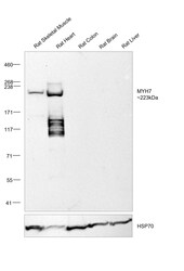
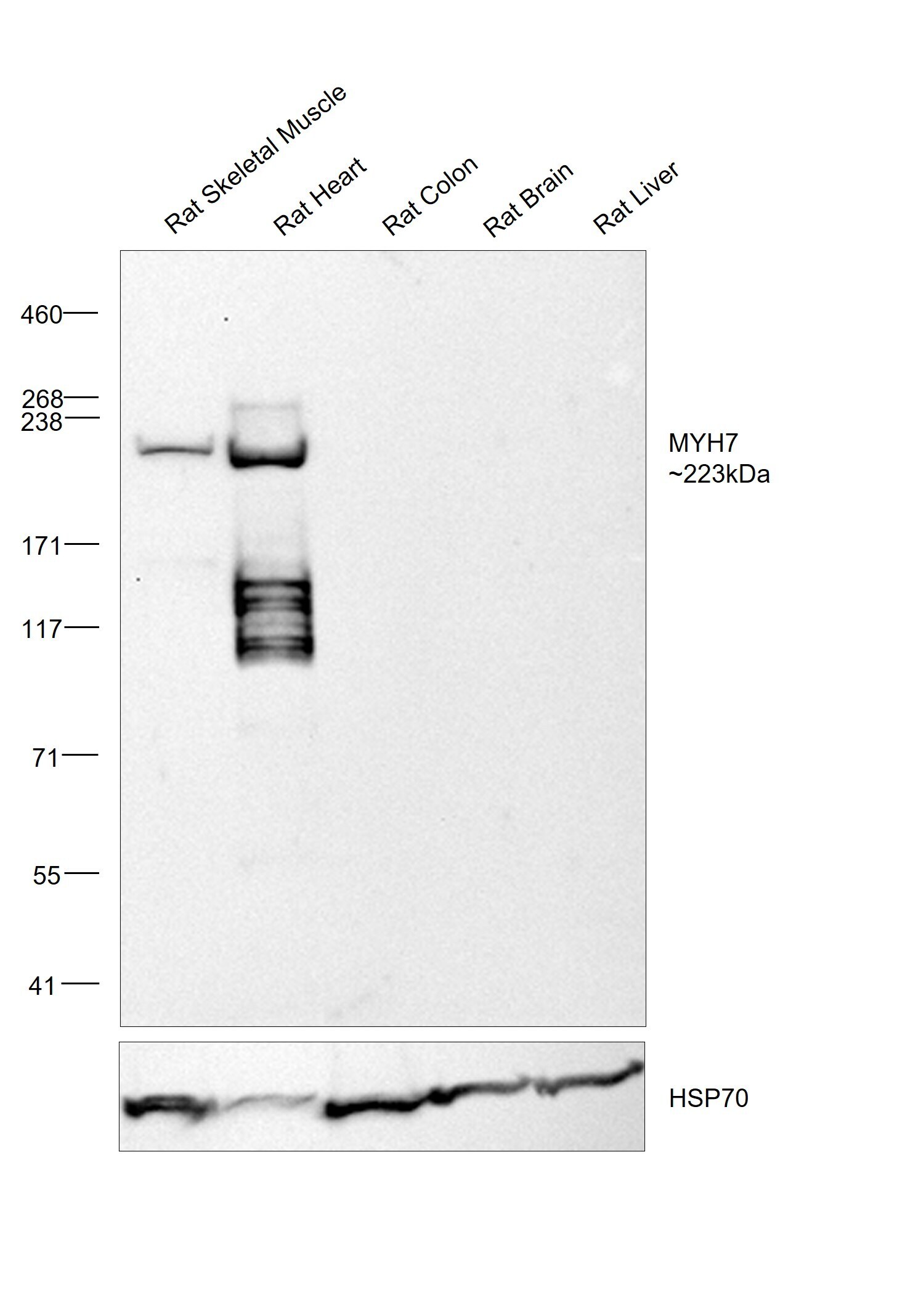
- Western blot was performed using MYH7 Monoclonal Antibody (NOQ7.5.4D) (Product # MA5-32986) and a 223 kDa band corresponding to MYH7 was observed in Rat Skeletal Muscle and Rat Heart (cardiac muscle) but not in Rat Colon (smooth muscle), Rat Brain and Rat Liver. Tissue extracts (30 µg lysate) of Rat Skeletal Muscle (Lane 1), Rat Heart (Lane 2), Rat Colon (Lane 3), Rat Brain (Lane 4) and Rat Liver (Lane 5) were electrophoresed using NuPAGE™ 3-8% Tris-Acetate Protein Gel (Product # EA0378BOX),10 well. Resolved proteins were then transferred onto a nitrocellulose membrane (Product # IB23001) by iBlot® 2 Dry Blotting System (Product # IB21001). The blot was probed with the primary antibody (0.1 µg/mL) and detected by chemiluminescence with Goat anti-Mouse IgG (H+L) Superclonal™ Recombinant Secondary Antibody, HRP (Product # A28177, 1:20,000 dilution) using the iBright™ FL1500 Imaging System (Product # A44115). Chemiluminescent detection was performed using SuperSignal™ West Pico PLUS Chemiluminescent Substrate (Product # 34580).
- Submitted by
- Invitrogen Antibodies (provider)
- Main image
- Experimental details
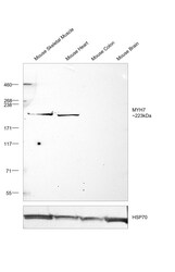
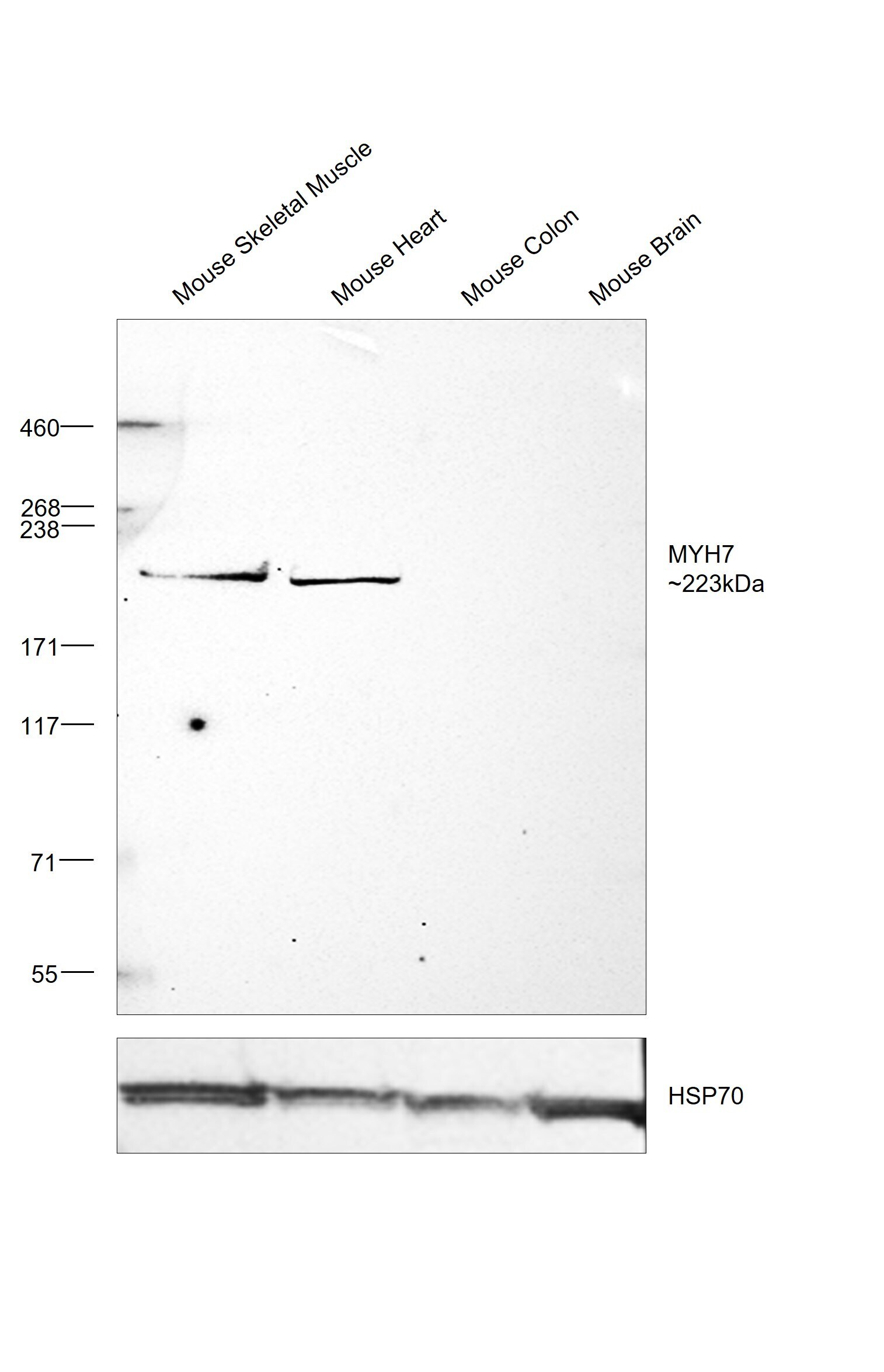
- Western blot was performed using MYH7 Monoclonal Antibody (NOQ7.5.4D) (Product # MA5-32986) and a 223 kDa band corresponding to MYH7 was observed in Mouse Skeletal Muscle and Mouse Heart (cardiac muscle) but not in Mouse Colon (smooth muscle) and Mouse Brain. Tissue extracts (30 µg lysate) of Mouse Skeletal Muscle (Lane 1), Mouse Heart (Lane 2), Mouse Colon (Lane 3) and Mouse Brain (Lane 4) were electrophoresed using NuPAGE™ 3-8% Tris-Acetate Protein Gel (Product # EA0378BOX). Resolved proteins were then transferred onto a nitrocellulose membrane (Product # IB23002) by iBlot® 2 Dry Blotting System (Product # IB21001). The blot was probed with the primary antibody (1:1,000) and detected by chemiluminescence with Goat anti-Mouse IgG (H+L) Superclonal™ Recombinant Secondary Antibody, HRP (Product # A28177, 1:20,000) using the iBright™ FL1500 Imaging System (Product # A44115). Chemiluminescent detection was performed using SuperSignal™ West Pico PLUS Chemiluminescent Substrate (Product # 34580).
- Submitted by
- Invitrogen Antibodies (provider)
- Main image
- Experimental details
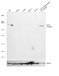
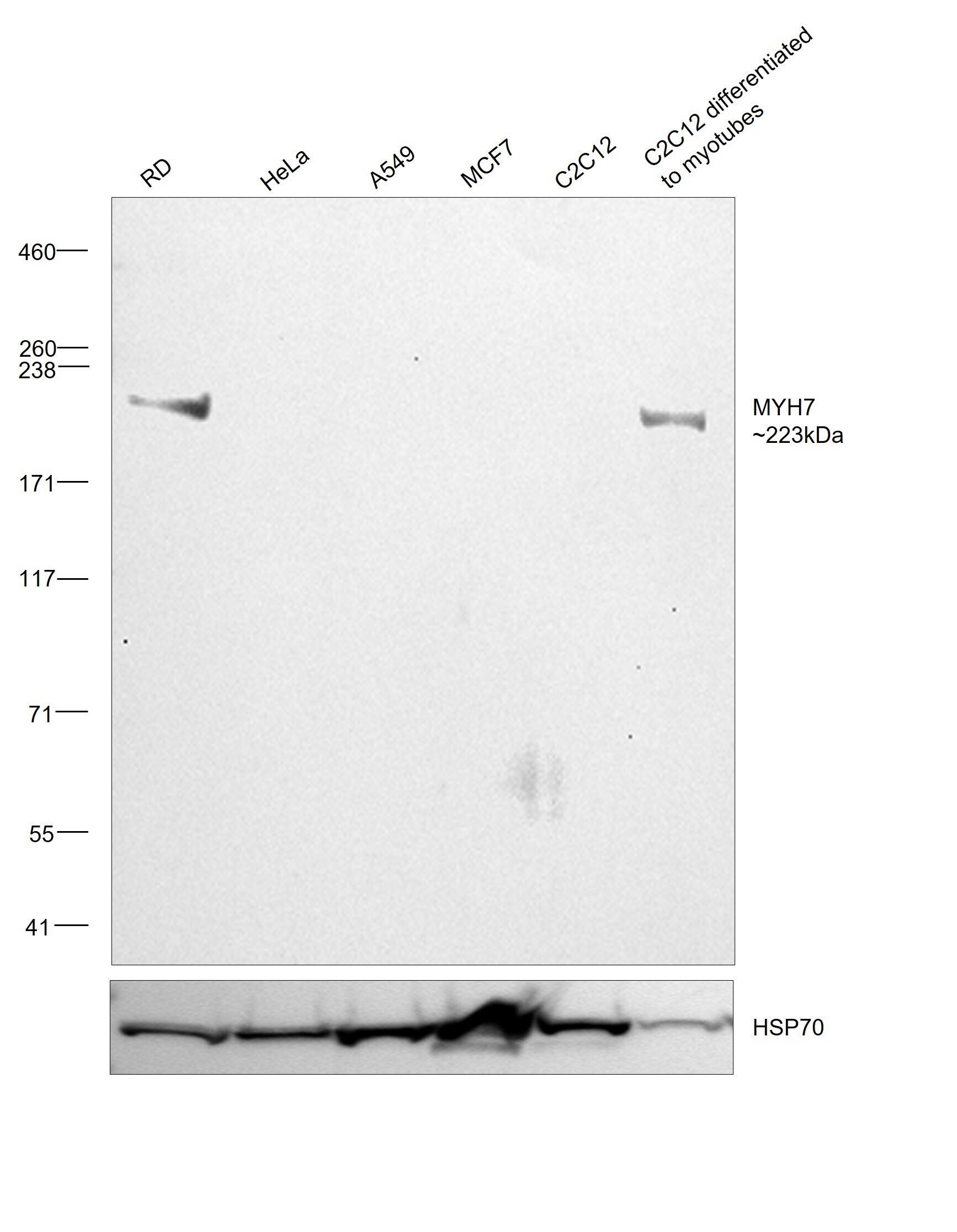
- Western blot was performed using MYH7 Monoclonal Antibody (NOQ7.5.4D) (Product # MA5-32986) and a 223 kDa band corresponding to MYH7 was observed in RD and C2C12 differentiated to myotubes but not in HeLa, A549, MCF7 and C2C12. Whole cell extracts (30 µg lysate) of RD (Lane 1), HeLa (Lane 2), A549 (Lane 3), MCF7 (Lane 4), C2C12 (Lane 5) and C2C12 differentiated to myotubes (Lane 6) were electrophoresed using NuPAGE™ 3-8% Tris-Acetate Protein Gel (Product # EA0378BOX), 10 well. Resolved proteins were then transferred onto a nitrocellulose membrane (Product # IB23001) by iBlot® 2 Dry Blotting System (Product # IB21001). The blot was probed with the primary antibody (1:1,000) and detected by chemiluminescence with Goat anti-Mouse IgG (H+L) Superclonal™ Recombinant Secondary Antibody, HRP (Product # A28177, 1:20,000) using the iBright™ FL1500 Imaging System (Product # A44115). Chemiluminescent detection was performed using SuperSignal™ West Pico PLUS Chemiluminescent Substrate (Product # 34580).
- Submitted by
- Invitrogen Antibodies (provider)
- Main image
- Experimental details
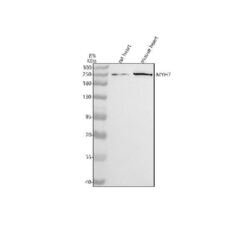
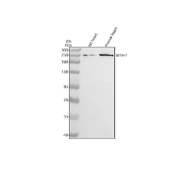
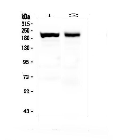
- Western blot analysis of MYH7 using MYH7 Monoclonal Antibody (NOQ7.5.4D) (Product # MA5-32986). Electrophoresis was performed on a 5-20% SDS-PAGE gel at 70V (Stacking gel)/90V (Resolving gel) for 2-3 hours. The sample well of each lane was loaded with 30 µg of sample under reducing conditions. Lane 1: rat heart tissue lysates. Lane 2: mouse heart tissue lysates. After electrophoresis, proteins were transferred to a nitrocellulose membrane at 150 mA for 50-90 minutes. Blocked the membrane with 5% non-fat milk/TBS for 1.5 hour at RT. The membrane was incubated with MYH7 Monoclonal Antibody (NOQ7.5.4D) at 0.5 µg/mL overnight at 4°C, then washed with TBS-0.1%Tween 3 times with 5 minutes each and probed with a goat anti-mouse IgG-HRP secondary antibody at a dilution of 1:5,000 for 1.5 hour at RT. The signal is developed using an Enhanced Chemiluminescent detection (ECL) kit with Tanon 5200 system. A specific band was detected for MYH7 at approximately 223 kDa. The expected band size for MYH7 is at 223 kDa.
- Submitted by
- Invitrogen Antibodies (provider)
- Main image
- Experimental details
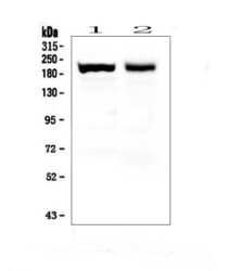
- Western blot analysis of MYH7 in Lane 1: mouse skeletal muscle tissue lysates, Lane 2: rat skeletal muscle tissue lysates. Electrophoresis was performed with 5-20% SDS-PAGE gel (70V, Stacking gel; 90V Resolving gel, Time: 2-3 hours), transferred to a nitrocellulose membrane and blocked using 5% Non-fat Milk/TBS (1.5 hrs at room temperature). Samples were incubated with MYH7 monoclonal antibody (Product # MA5-32986) using a 0.5 µg/mL dilution, followed by a goat anti-mouse IgG-HRP at a dilution of 1:10,000, and developed with enhanced chemiluminescence (ECL).
Supportive validation
- Submitted by
- Invitrogen Antibodies (provider)
- Main image
- Experimental details
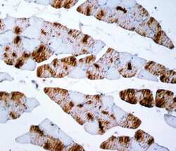
- Immunohistochemistry (Paraffin) analysis of MYH7 in paraffin-embedded section of rat skeletal muscle tissue using MYH7 Monoclonal Antibody (NOQ7.5.4D) (Product # MA5-32986). Heat mediated antigen retrieval was performed in EDTA buffer (pH 8.0, epitope retrieval solution). The tissue section was blocked with 10% goat serum. The tissue section was then incubated with the primary antibody at a 2 µg/mL dilution overnight at 4°C. Peroxidase conjugated goat anti-mouse IgG was used as secondary antibody and incubated for 30 minutes at 37°C. The tissue section was developed using HRP Conjugated Mouse IgG Super Vision Assay Kit with DAB as the chromogen.
